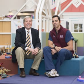
MUSC Children's Health pediatric neurosurgeon Ramin Eskandari, M.D ., and MUSC Health craniofacial surgeon Jason P. Ulm, M.D . discuss surgical approaches to correct craniosynostosis or the premature fusion of one or more of the brain’s sutures. Also view the companion video in which Dr. Eskandari provides commentary on surgical photographs from a recent case.
RAMIN ESKANDARI: Craniosynostosis is a condition in which the bones of the infant skull fuse abnormally. And usually, when we say abnormal, it means early. That fusion can happen before they're even born. And so a child is born with craniosynostosis, or craniosynostosis can develop shortly after birth, as the bones don't tend to splay apart from each other appropriately and so they fuse early. There's lots of different types of craniosynostosis. By definition, any of the bones of the skull that prematurely or abnormally fuse can be called craniosynostosis. The most common form is called sagittal craniosynostosis and that's when the suture that's right in the middle, connecting the two halves of the bone on the parietal bones, fuse early. And that's the most common one. It gives you a head shape that's called scaphocephalic. It's elongated, sort of football-shaped. The incidence of that has been reported in the one in every 5,000 range in the number of live births. Depending on statistics you look at, the next most common could be considered metopic craniosynostosis or unicoronal. Metopic is the one that's right down the forehead, and it gives you a triangle-shaped head. And the coronal one is the suture that goes across like a headband. The most common form of that is one side being fused. Having both sides fused is actually a little less common. And the incidence in those cases is one in 7,000 to one in 10,000, depending on which suture you're talking about. JASON P. ULM: So, craniosynostosis differs from head-shape deformities, or deformational plagiocephaly, in that the latter is not a fusion of the suture itself but rather pressure from the child laying in one particular way or another on the bed. The Back to Sleep Campaign has led to an increased incidence of these cases. And the treatment for such is different. Whereas, with deformational plagiocephaly, it's more an education for the family and waiting for the child to grow where he's not laying, or she not laying, on their back side, and they're turning over more, sitting more upright. Oftentimes, the head corrects its shape in itself. In severe forms, we may recommend a helmet orthotic therapy to help normalize the head shape. In craniosynostosis, if there is closure of the suture, that requires, more often than not, surgical opening of that suture, as well as potentially reshaping of the cranial vault. RAMIN ESKANDARI: The surgical goals for any kid that has craniosynostosis is pretty much twofold. Number one, release the abnormal suture. So take out the area of bone that has been fused, create a false suture, basically. And then number two, to try and get some element of expansion of the skull in a direction that you're going to be able to rely on as the brain grows. So, for example, when you have synostosis of the sagittal suture, the first thing we do is we remove that area of the bone. And the second thing we do is we cut what we call barrel stave osteotomy-- it's basically like a blooming onion-- cut that into the skull so that, when you're finished with surgery, as the brain grows it wants to grow sideways, preferentially, instead of growing more in a forward-backward direction. The more complex cases of craniosynostosis, especially those that have multiple sutures involved, sometimes require more extensive surgery. And that can be a staged surgery. Sometimes, when you have complex synostosis in a kid who is very affected super early in life, where they don't have a lot of bone to work with, you sometimes stage the operation. So you do one part when they're early, three to six months, and then another part maybe a year after that. And in those cases, you're remodeling the cranial vault, the head. And that remodeling can involve as extensive as complete removal of the entire cranial vault and reshaping it and cutting it into pieces, or taking parts of the skull that are already there, that are normal, slicing them in half like an Oreo, thickness-wise, and then using that extra bone to remodel the head shape. So there's lots of different options. This is one of those disease processes where there's no one right way to do it but several possibilities. JASON P. ULM: So, at MUSC, we perform between 20 and 40 craniosynostosis procedures a year, sometimes more. So I would characterize that as a high-volume center. And it's important for patients to go to high-volume centers where they have the expertise and the surrounding skill set of the team involved that is used to and capable of taking care of the complexity of these cases. RAMIN ESKANDARI: The method by which we're helping these kids here is a method that, I think, a lot of multi-universities, high-level universities, do it, is a multi-disciplinary approach. So, here we have myself, my partner, who is another pediatric neurosurgeon, and then we have Dr. Ulm, who is a pediatric plastic surgeon trained in craniofacial plastics. And we all work together. So, usually it involves at least one pediatric neurosurgeon, one pediatric plastic craniofacial surgeon, and then the team-- the way that we do it here-- the team of engineers that helps to reconstruct the skulls for us. JASON P. ULM: So, 3D printing is a new innovation that has been used in the past several years. In particular, it's being used by us for modeling of the skulls, for craniosynostosis surgery, which gives us an in-hand model that allows us to design and simulate the cuts that we would be making during the operative procedure. RAMIN ESKANDARI: I think that part is a little bit different. Not a lot of places have that as an option. We tend to use that on almost every case nowadays because we're finding that, having that 3D-printed model, we can see all the aspects of surgery, and we can even practice the surgery before we even get in there. And it's that patient's skull. It's not like we're doing this off of a computer. We physically have our hands on it. We can rotate the skull. We can look on the inside of the skull. We can anticipate any areas of bony abnormality that we never would have known beforehand. And we go into the surgery having a plan in place that we both agree on before surgery. And we can share that with the families as well. My takeaway message for craniosynostosis, I think, is don't be afraid to ask a specialist for an opinion. A lot of times, we can try and diagnose either the type of craniosynostosis or reassure a family that it's really not, that it's more positional. Just by seeing the patient, there's a lot of nuances to the way their head shape looks-- the forehead, the eye levels, the ears-- that might require someone who's seen it a few times to be able to pick up on it. And secondarily, the imaging that we get is very different, depending on the age of the patient and what we're actually trying to achieve. So seeing us initially might not be a bad way to go because we can defer some imaging that they might not need. Or we can get imaging specific to their condition that we would need for surgery.
Related Presenters

