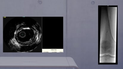
This is a recorded live case focused on the multi-specialty approach to the treatment of patients with venous disease. This recorded live case will feature dialog on the overall evaluation of the vein patient, how to treat patients with both superficial and deep disease and non-thrombotic and post-thrombotic lesions.
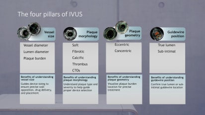
I have the pleasure of introducing our faculty today. So we have dr steve Elias who's a vascular surgeon from Englewood Hospital in Englewood New Jersey. He is the director of center of the vein disease at Englewood Hospital. He is also the founder and course director of expert expert venus management. Dr Elias is also the medical director of vein magazine and medical editor of the american venus forums, newsletter vein specialists. We also have the pleasure of having doctors anna Safadi from um ST mary's Medical Center in Hobart indiana. He is an interventional cardiologist and he has authored several articles on P. E. And his practice has grown tremendously in the area of deep vein intervention particularly in the way of venus compression compression syndrome and treatment of DVT. He is here to moderate the case. So welcome thank you doctor the body and thank you dr Elias and dr Elias I'll hand it over to you so you can tell us about today's patient. Okay good morning everybody and uh glad you're here. I'll tell you a little bit about the patient. We've done some preliminary work already to get access and everything else but we'll talk to it. But this patient is 60 year old male who does have a history of a previous DVT said to be only in his uh in for England venus system. He has some scarring in there when we imaged by ultrasound. But he also has some mild post robotic disease and thickening of his iliac veins and also some compression. Which we're going to show you all of this. He work standing up the great majority of the day and I actually saw him over a year ago. He's from east of west of here in pennsylvania. Uh And it was just his symptoms bothered him enough in that he could not work a full day. His leg was very swollen and um he really couldn't tolerate compression stockings. And that's that's kind of a key thing. When you ask patients who are post storm biotics or have some type of compression, They can't tolerate compression stockings. It's because when you're trying to put the stocking and trying to squeeze that blood back up towards the heart, it's meeting and obstruction. So you're hitting your you're squeezing against a a firm leg that cannot empty. As opposed to people have insufficiency, they will say to you, yeah, when I wear the stockings, I feel better because their problem is not obstruction, its insufficiency. So that's his story as what we would call kind of venus qualification. I did a back in july end of july, a diagnostic ivascyn venogram, because I like to sit down and talk to the patient and tell them what they have and then he decided to come back. So, in terms of the procedure itself, what we've already done, and if whoever's working the camera can hone in on the groin area over here. Okay, We have made access. He had had an ablation in the past at another facility of his great staff in his vein for the same symptoms that he has now. And he said those symptoms did not get any better because clearly a big swollen leg uh with venus qualification is not being caused by an incompetent great stuff in the same. So we used the stump of that vein to access into the deep venous system. And access points. People can use, I like the Great staff. And if it's there, you can use the femoral vein in the upper third of the thigh as well. He's a little chubby kind of guy and luckily were able to get access into that stuff of the staff. And the important thing is you don't in general want to access the common femoral vein because when you put your eye this up and it's going to stand or not, when you put your sheath in, you maybe halfway into the external iliac vein already and not see disease that is in the external iliac vein because the sheep has gone above that. So you like to kind of keep your sheet within your common thermal vein one way or the other by making access in the cevennes or by making access in the upper third of the fight in the femoral vein. And so here you can see we've made the access uh in that stump or in the upper third of the thigh we have a 10 French Sheath introducer because that allows us to both. You see, I've this device which goes to an eight French and also any balloons or stents that we may uh need most. Most stents will fit uh attend into a tent. Some will be smaller but we've accessed with the tent so we can use whatever size we really need. Then um we've done a hmd venogram and Phil can you can you run can we do that run? All right. We can do it right here. Can you guys show us the uh flores coptic screen and we're going to show you guys a run from our what we did previous of just the hmd venogram. Yeah. There you go. Thank you. Yeah. So here's our snd venogram. We wound up pulling the sheet back because we're a little too far in do the second. So here you can see external iliac common iliac which looks a little smaller than the external iliac and the I. V. C. Okay okay let's go to the next run nick. Can you do the next one? So we pulled the sheet back a little bit and we came down a little bit to look lower down. Now it's higher that's what we got. Yeah. Okay this is our last run here. So again you can see there's narrowing the venture. Get mhm bigger as you go up. But you can see that the vein the external iliac vein below is larger than the common iliac vein where it meets the vena cava. So that gave us an idea there was a problem then we did some I've just runs which we're going to show you live now instead what we do before we do anything else is we get a long wire up and we have a 035 amplats wire going from the access site all the way up into the right internal jugular vein. And the reason we do that is if the stent, God forbid it starts to float and it's not going to wind up in the heart, at least you have control along the whole venus system so that it doesn't get stuck within the heart itself. And that's why we do that. It may take a little bit of time today. It was relatively simple, but I think it's well worth doing. Uh, so that you feel total control and you don't have to worry as much if the whatever reason the stent, when you deploy, it starts migrating towards the heart. So that's what we have in here. We have uh the offices over that wire. And now we're going to show you a uh live by this image and then we'll show you some of the images we took before. So, nick if you could hold the wire and when we're doing this. And most of you who have catheter skills know that your wire is your guide to everything. If you lose access. And the whole case takes much much longer. It's a big pain in the neck. So nica vascular fellow is holding the wire down down below uh by the ankle area. And then I'm going to go up with the service. We've also sewn in the 10 French sheep because when you're exchanging back and forth, you don't want to pull the sheet out. So, I always so the sheath in place as well. All right. So, if you look at the Ibis here, Mhm. And I'm going to be looking at the Ibis myself. So, we're going up and here's here's a common formal. Then you can see a little bit of scar tissue on the left side of the screen and it's a fairly large common thermal vein. We're going up first, we keep going up is external iliac vein. And you're going to see soon the area of compression as a right external and internal really like arteries divide and here they are compressing the vein going up. Now we're in common iliac thing, which again, you have some idea there's some thickening of the wall, but as we go first, see how it gets much narrower and much thicker. We're right at the bifurcation. Now, if you look around the 10 o'clock position, that's the confidence, that's the left common iliac vein uh and the right common iliac vein with a catheter in And if I go up higher, we're now in the Vienna Kaveh itself. So it's important for you to understand the images. So when vena cava, now we're coming down, you're going to see the bifurcation as we pull down and slowly pulling down. So we're pulling down and here you see the vindictive is beginning to divide into the right and left. There is a good example. Nine o'clock is the left common iliac vein and in the center is the right common iliac vein, which is thickened and somewhat small. So we're going to lay out stent, I'm going to go up a little bit, a little bit into the vena cava kind of right here and then we're going to come down. This is a thickened uh vein much smaller than it's supposed to be. And we'll show you the images of what that is uh, that we took measurements before. So here's common iliac vein still on the right, I'm still coming down. This is the right leg we're working on and this is the take off of the internal iliac vein. You can see around the two o'clock position I'm going up and down. So Here is that out budding at the 2:00 position. We come down now in external iliac vein and here's compression starting and you come down as you come below the area of compression, you're going to see. The common thermal vein is quite large and just allow extra Elliot Krane. And now we're coming down to the common thermal vein which you can see here is very very large. And obviously the common thermal is supposed to be smaller than the external and the extra is supposed to be smaller than the common iliac thing. So now if we can go uh to the images we took before. Yeah. Yeah. Mhm. We'll show you some of the measurements that we took 44 So here in green is our compression area Which we measured at 83 sq mm. And the reference area below is 206 square millimeters. Which is is somewhat dilated in the external iliac vein. It's supposed to be about 1 50 square millimeters plus or minus 10%. To get debilitation debilitation below the area where the stenosis is because clearly it's going up. Uh and your media resistance of the vein below dilates. And the comment, I think we have a measurement of the common central vein as well. Let's see what that is to show you know. Mhm. Right. This is the extent of this measurement. Here is the external iliac vein kind of uh midway between where we have compression and the common iliac. And that you can see measures 211 sq mm. But also you use the diameters which you can see on the bottom left Of 18 and 14. If you add that together, that's 32 and then you divide by 2.5 is 16 and that gives you the 16 millimeter diameter is the extent that we're going to use, we're using the vinovo stent today. So it's a dedicated venus stent. And when it says it's 16, it is 16 in the newest sense. We don't really want to oversize. Uh, in the old days when we're using wall sent only, we would oversize because by, by two millimeters. But here, if you oversize, patients will get some significant pain because you're really stretching that that thing. So we're gonna use this, we're going to go with the 16 but show us the image of the common. This is again, leave that we've got a this is a little bit lower down on the external. And again, you can see we're in the range of a 16 and maybe a little bit smaller. But I would like you want the stent to sit in place so you don't want to undersize because you don't want floating. You wanted to sit in place in the venus system opposed to the venus. Whoa. And that will come down to the common federal. Which we should have. That's another You should have the last one. Yeah, right there, nope. Well, mm we're just looking for the common formal, here we are. So our common fellow vein measures 289 square millimeters, which is way bigger than it's supposed to be. It's supposed to be about 1 25 or so. So again, it's because of the proximal obstruction. He has really two areas. He has the area in the external iliac vein where the arteries are compressing and then he has that post robotic area higher up with the thick and uh they want I just asked before I go on any questions come in at all from anybody yet. Uh steve, I wanna uh highlight a couple points. Good morning. How are you? I think it's important to um really um uh explain to the audience the the use of a stiff AM plus type wire. Um I've seen people use the standard J wires and especially if you're accessing the proximal femoral vein or even sometimes the gsp. Uh sometimes these these veins are a little bit deeper and if you don't use a stiff wire you will kink the sheet and then you will be really kicking yourself trying to figure out how to deal with that later. So, unlike other types of uh lesions that we go after, it's very important to do the stiff wire and anchor it high. Like you mentioned to really have a strong rail and have safety profiles um just in case you get into issues. Uh number two I think um you very clearly showed nicely the confluence. Um and when operators are doing this very important to really um kind of flora saved the tip of the ibis catheter to a bony landmarks. So you're very, very accurate and you can be very accurate as steve is going to show that placement where to put that stent. Uh And then thirdly, as you mentioned, to highlight that you really need to size the stent um based on that external iliac vein that you showed, because if you go to the priest cyanotic areas, you're gonna oversize and cause a lot of problems to the patient. So I just want to highlight those points because I've seen people not do that properly and get into issue. Right Okay. No thank you and thank you for highlighting that. So question yes. Allies, yep. Do you worry about that thick vein while not expanding completely? So we're going to pre dilate before we we placed the stent. So we're gonna we're gonna use a 16 balloon to pre-dilate that area and stretch it a little bit. And then the stents themselves of a noble extent is very very strong. It will it will stretch that vein to just about 16 millimeter diameter. So yes we worry about it but we will pre dilate especially in patients who have if they had a pure compression and never had a DVT and not a tick in vain. Some people do not pre dilate, they'll just lay the sent in and it will usually be fine. But in this case we're gonna pre dilate any other questions. That's also an important point. So for thrombosis patients, um uh it's very important to pre dilate. There's a lot of fibrosis, there's webbing sometimes that you don't really appreciate. Uh and you should generally pre dilate 1 to 1 or maybe two millimeters smaller but to really expand and see how the lesion behave, particularly in the pram biotic patient. Because sometimes if you go in direct stent you, it actually may pop the steps out or cause a problem in terms of accuracy of placement. So I think that's also very important to kind of just highlight to everybody that the concept of pre dilating in the probiotic patient right now. Uh we're going to show you on the flora screen how we decide where to where our landing zone is going to be. So if you can give us the floral as well guys floor and I this nick, hold the wire. Yeah, let's go. Ok, so I'm going to go live with the flow. Okay, You're going to see the I'm going to place the the Washington. I'm going to place the ibis go live please with Agnes. Here we go. So we're going up with Davis. Going up, here's a conference right here. Okay, this is vina Kaveh. And I'm coming down to confidence and we're going to want to go just above this conference because there is some scarring in here. So we're probably gonna land out. Spent about a half a centimeter into the uh vindicated. Mhm. And this is kind of going to be our landing zone. Now, if you look on the screen, you cannot see what we have done already at the tip of this ibis catheter on our floor. Oh screen. We have drawn that. This is where we want our landing zone to be. So you can either use a bony landmark or we can use um the marking. So what you can see here is we have the letter L. Next you can point on there. I don't know if you can. No. The letter L nick is here. That's going to be a proximal landing zone. Or as we said last night, our cranial landing zone and the sea right below that is where that compression starts in the common iliac. Then we saw the thickened area. Then uh if you come down way down to the mid portion where we say oom, that's the compression where the iliac veins are squeezing on the arteries are squeezing on the vein. And then the last one is our landing zone. But actually we're probably gonna wind up way down here at our landing zone. Uh That's what we decided we where we want to go. And if you count there's each, the ibis has centimeter markings. We counted. It is a length of 16 cm. So we're going to use a stent. That's gonna be a 16 by 1 60 millimeter length stent. Mhm. And that's what's really nice about the service is you can not only see the pathology from the inside, but we use this as a way of exactly marking where we want to land both cranial and uh quartet. So honestly have any remarks about this. No, that's very important point. So it's important to understand the to venus stents on the market. There are some differences in terms of both sides, diameter and length. So when you do measurements like that it will facilitate also selection of stent. So for example, the V. C. The boston scientific fact The biggest diameter is 16 and the longest diameter is 120. Uh And that's a nine firm system whereas the vinovo stent um you can get up to 20 millimeters and you can go as long as 1 60. So in a lesion like this where you want uh the uh you know where you really want to minimize the number of stents you can get away with one long stent being a 1 60 then a diameter of 16. And of course you would have to put a 10 different system for the venerable if it's 16 or more. Uh So it's extremely I mean when I hear people um you know say they do venus intervention without I've I tell them you know where I say to the person who I hear from, its because there is no way in the world you would be able to size appropriately. You will be able to delineate clearly. Like steve has on this image placement of the cranial and caudal, synthesize the distribution, the location of compression. So I this is absolutely essential to doing appropriate the dvt and venus. And uh it really shows it really nicely. All right guys, thank you very much. So we're going to go back. So this is how we measured it. This is what we can come up with again. You can look on the screen, we have measured everything and we are now on the I. V. Screen. We're right at our landing zone above again we're going to come back down, remember we're going to show you once again this is the area that's somewhat thickened here coming down in the common iliac vein. Now you see it's not so bad. The vein wall is not all that white. But if you come up here you can see there's a thickened area. So you can see how much you can see with avis. And then there's our area of the compression and we'll come back down. And now what we're going to do is we're going to pre dilate. So we're gonna use our 16 billion with 16 by what, 60? Perfect. 16 x 16 balloon. Every time we go in and out I say tunic vascular fellow I have wire, he tells me he is why when we go in and we wipe down the wire you can take the i this image off now and just have an image of us live. And the chloral image based on I've is what was the reason you decided to extend the sent down to the kind of mid common femoral vein? Well it's not it's really distal external iliac vein. The reason we did was uh the area in between uh some of them measured a little more than 16. And I really like to send to sit in nicely. So yeah, I mean if you want me to show you again, I'll show you. Let's just I just want to clarify just why not? Everybody should, we'll take another three minutes. We're gonna put the ideas back up again and I'll show you. Yeah louis just take this care so we don't lose it. Perfect. So put the harvest back up. That's a good question. The question is, where do you land your Kordell landing zone? And first of all, as we spoke about last night, I don't like to land deep in the pelvis. So I'd like to come around the curve of the of the pelvis and into the external iliac vein. Can good. They have the I this image up again. So now I'm going to go up again and I'm gonna show you I have to look on the floor. It's been I'm gonna Uh huh. So this was a question where we are now. This was a question of our landing zone. And Maureen, can you go measure? So this we thought perhaps but I'm gonna show you it still looks a little dilated compared to let me come down first. Hang on, Maureen, go live. Compared to here. You see how it's a little smaller here, honest. Mm Compared to here, If I go up, see this is still fairly large. Uh mainly to anchor it. Yeah, for the side, right, It's angering. And we're above the common criminal vein. Here, we're not in the common summer. Just so here she is measuring on the screen the area that I thought it was a little big to land. So that becomes a say 19 plus 16 is 35. So it's a little bigger than a 16 stent. I didn't feel comfortable, but I don't want the stent to float, right? You want this tend to to shit. So that's why you go live and I'm gonna come live with my flow. That's why if you come down here where we think we're gonna and you can see where I've asses. Now we measure this vein. You see it's smaller. Yeah. And Maureen is measuring it. It's a very good question because the worst thing you want to do is not have to stand sit in place and move. So this year is measures like 29 30, so that's going to sit in 16 is going to sit in very well there. You know what I'm saying? Very good point. I all that. I think it's also important to highlight to the audience the concept of, um if you, let's say for example, you need to extend the sentence slower and a lot of people who are not comfortable with being a stenting and or more on the arterial side, they're not comfortable putting sent across England ligament obviously. But in the venus world, as we talked about last night, particularly in the Rombach ticks, um, you need open to open. So, um, there is plenty of data showing safety of scenting across the England malignant. If you need to extend even all the way down to the lesser trow cantor distribution, above the propaganda. Uh, but I think if you're gonna do that, I was once again is extremely important because you want to place the initial stent if you don't have one to go all the way down uh in an area where the overlap will not be kind of in that England a ligament distribution. That's really important. You don't wanna double layer. Uh I think uh in that distribution because it could potentially cause problems. That overlap zone needs to be almost where you're where you know where your initial potential landing zone, not necessarily for this case, but I just want to highlight the concept of if you're going to overlap stents, try to avoid overlapping in that area. Kind of a traditional external common criminal vein distribution. And the I this will help you plan on size because you'll have the cm and you'll know where to land the initial sent overlap 1-2 cm and land the distal sent in a situation where that's needed. Yeah, good point, good point. So now we're we can just give us just a flow image. We don't need anything else. We're gonna put our balloon up uh and and dilate. So you have wire right now. Okay. And I just want you guys to know uh you know Nick is really giving up his experience here so you all can learn by because he's not going to do too much of this case here. Of course. So can you bring the floor screen closer to me please? So you can see here with well coming up without balloon. Now you don't have the markings. Thank you martin. You don't have the markings that we have. Um But if you trust me here, where we are now is who we start. The top dot is at our uh landing zone and the bottom dot is just down the vein. So now we're going to go in here. Can you go Yeah go ahead. So Nick's gonna start to dilate this. Uh We're gonna floor oh you see the balloon is distended here keep going and this patient you can see a little bit of wasting their a little bit And we just leave it up for about 30 seconds or so. We don't need that much. We've given the patients 6000 of Hepburn before we just came on on air here. So obviously I have to be jeopardized. You tell whoever is giving some of the sedation or whatever an aesthetic thing. I do not do these people under awake. Some people do. I don't I don't want them to move. Once you have your measurements. If they move you have to re measure everything. So anesthesiologists are doing a good job here. Come on down. So now we're gonna come down with the balloon and we're gonna dilate lower down. I think we just have to dilate one more segment because after that everything is pretty normal. Some comments about technique as well. Um What antique regulation? I'm assuming you just standard heparin. And if so. What a ct. Just for people who are kind of getting into this. Well, I personally, I'm not doing a cts honestly, because it's a relatively short case and we just give him a bolas. I mean, some people do it. You do a ct scan it. Uh We took we have a machine there in the lab. It's very quick turnaround. But I've definitely done cases without it when things are not happening. Right? So, you know, um just uh just kind of kind of throwing that out for people just to be aware that you can do it in different ways. Um There's a question while you are where you well, you're okay. Well, you ask the question, wait honest, ask the second while you're asking the question we're going to uh well just direct the second segment but go ahead and go ahead. Yeah. So one of the attendees mentioned, you know, do you consider referencing standard sizes from the literature? Like dr Raju's articles out there in regards to standard, general size and you commented a little earlier, Like, you know, the comedy like vein is usually 1 75 to 200 millimeters square. The external iliac vein is usually 1 50 give or take, uh 1 75. And so if you're not able to necessarily, particularly like in an acute DVT, um, understand the uh, from Ivan, the actual size. Do you typically just use standard literature to help you and for reference sizing? And I think that's a good point. I often will use it if I cant success not in a case like this. This case is very clear. You can clearly see the size that you need. I think these standards as we're doing this on the screen, you'll see the wasting exactly where the compression is here. Absolutely. That's a good very nice picture of that. Okay so go ahead keep talking as I'm sorry we're going back and forth. So I no no go ahead. And so I do most of my cases with moderate sedation actually. So this would be a situation where I would warn the patient knowing where I know the compression and right where before we're going to pre dilate that they are going to have back pain. And we talked a little bit about this yesterday if you educate and inform the patient and try to and I will kind of wink at the nurse with sedation that it's time to be a little bit aggressive with sedation. Um But this is the time to do it because that leads in that waste there when you start pre dilating that it's going to hurt. Now obviously this patients under general anesthesia so it's a different situation but it's I think it's it's important whether you you know if you decide to do them under moderate you know that that you that you really really um engage the patient and make sure they know what to expect. Right? You have a big syringe into the second movement. Okay that's there was a good point. Also point I want to make is uh we don't have to pan the whole room here but this is a team effort when I do this it's not like I think I'm the doctor and I know everything and nobody else knows anything. So before we we placed the stent, I'm asking everybody who has ever run in the eye this machine, what do you think we should do? I'm asking the uh representative from uh Bard, whose makes uh vinovo stent, what you think this is the right size. Um And we triple check everything. And I say to anybody, if you have any thought that we're not doing the right thing, please tell me I don't. The last thing you want to do is make a mistake. So I think you need to use everybody's experience in the room. Uh And don't think you know everything and honest. If if you have any thoughts about doing something wrong, please jump right in. Okay now, so far so far it's going all right now, can you show okay? What I want to show is we're going to be taking the balloon the out. So Nick whose hand is up by the sheet, the 10 friend sheet is holding the sheet. I am aspirating With a 60cc syringe here and holding it as I told the balloon out. So I'm collapsing the balloon completely. Because what you don't want to do is pull out the whole sheet because you may meet a little resistance. So that's all we're doing. He's going to tell me when the balloon comes out and here it is, and now we can let go minor point. But you know, if you don't do that, sometimes you're gonna rip the whole sheet out of the patients, then you lose access again. I think that's a very important point. I think that's even more of an issue when you like, are using a smaller sheets like a nine french or even sometimes you can get away with Dave Friendship, you're doing a 12 millimeters then, and if you don't really go negative on that balloon blair's, you're gonna you're gonna you're gonna pull the sheets out if you don't know this. Pay attention. Yeah. So now we're ready to deploy the stent and we're gonna just opening the scent. I don't open up anything until I'm sure I needed meaning when we started the case here, all we had was a micro puncture set. And that was it because if I couldn't get access, I don't want to have, You know, uh stents open balloons, open everything else open. So it may take me another 5 to 10 minutes. But the last thing I want is to waste a lot of money, and especially if you're paying for it. So my suggestion is don't open up anything until you need it, because you don't want to waste waste money. So you can see here as I'm going in uh and nick is holding wire. Mhm. Right I'm going to go up and now we're gonna look at the floor image. Yeah. So you cannot see the markings that we showed you before. But where I have the stent right now is where we said we want our landing zone to be and you can see we're coming down a little lower than we thought. But that's okay about the santa meter. Uh I'm looking at my corner which you cannot see but I can see. So so I think I just want to bring up this point because I've had this issue come up with me as well where when you're looking at the right side and you use Ivan Sometimes the stent distantly will be slightly lower than you think it is, particularly compared to the left side because the left side of that external Iliac vein does a sharper curve. And um uh even when you drive it sometimes the disco aspect of not 100 where it's shown on I've it. Um And so it's important to note that also if you look at this you know under floor, he was showing you the vinovo stand um um you know where that distal marker is is where that stent is gonna land. Uh And it has a very very simple and he'll show us steve um deployment mechanism with with the rolling mechanism which is very nice and very accurate. Uh And also which is not that will say the actual size of the stent right there. You know just in case because I've seen with Walston sometimes people make mistakes because they don't write the size of the stent and you get the wrong one and you're not sure. And then it's a big headache. Right? Good point. So now you can, whoever's working the camera, you want to come in a little bit more on my hands and we'll show you this nice rolling mechanism here. Okay, you can see it here. There's a big wheel and a small wheel. The small wheel deploys the stent faster than the big wheel personally. I never used a small wheel. I like to extrude the stent once we see it flour, which you'll see on this on the screen, I then Pull it back to our landing zone, deploy about 1/3 of it. It's a nightingale stent, let it warm up, let it sit in place and then continue deployment. I don't just zip the whole thing out at once. So we'll do here. We're Now, this is kind of a crucial part we read is a locking mechanism so we're going to unlock it and we're going to go live again. And so that's very important. Unlike waltz sense. You know this you cannot receive the stent once you start deploying. Uh you know you're you're pretty much done so flower pull it down exactly where you want it and then kind of like steve mentioned, you know, deploy the rest of it slowly and and you place it for for so about a half a centimeter above where I want to be. So ultimately so I'm going to slowly deploy. Mhm Wait till the flowers here mm. Ben and everybody. When you see the flower you let me know community mhm started. Yeah. Mhm Yeah. Mhm. Okay. What? There we go. So here's our firing. I'm going to come down a little bit. Yeah right and now I'm keeping everything steady was slowly deploying. You can see the stent deploying here and now I'm just gonna wait I'm gonna let it warm up for about 30 seconds. Mhm. Yeah OK Walmart 30 seconds always seems longer. Yeah I am holding the white part of the stent is a try actual system. You can't hold the dark part because that's moving as you're deploying so we're gonna start to continue to deploy. So once again if the patients under moderate sedation this is the time to also warn them about back pain and possibly give him a little bit extra uh medication is needed because once especially goes right here where he's at now where that compression was most significant. Um Now in this case he's intubated and under general but if you do it under moderate they're going to feel it here yep. You're absolutely right. And um we're waiting again. So about two thirds of the stent has been extruded. Yeah. Like to warm up also you tell them they're gonna feel most of them will have some back discomfort post procedure as well. And if you tell them that beforehand they're okay with it. Okay? So hopefully now we'll just extrude the rest of the stent here. Okay? And so now we're extruded. Now when you go in and out of any stent I do it under the floor. I don't just pull the stent down now without looking at this. God forbid you want something to get caught on the stent and you'll just great. So now we're just gonna come back down and again nick is going to hold the sheep. Remember access is everything you lose access, you can't do the case. So I'm coming down slowly coming out fine here you're good nick, Yes, I'm going to have it so when it goes wire, okay now we're going to go before I shove a balloon up again. I'm just gonna look with IV's. I want to show you. Let's see how it looks and see if what areas we might have to do some. Perfect. We also were wiping the wire off with some Hepburn solution to keep any promise or nick. Reload the uh five ish. Do you always post dilate your stents, both robotic and levels Or do you not necessarily with nibbles? I may not with this one. I probably will but I want to identify there any particular areas I really want to make sure and areas I may not have to. So that's why I'm going up now before just dilating. Um Again, everybody is different. Some people just post highlight right now. Uh again, I'm going in to the stent. So I am looking with I'm looking with the Flora to make sure. So I'm going up now you can see on the Ivies image the stent is in place. We're going up we're going up and notice I'm not flowing. Now there's no reason to use excess radiation if I can see on I. V. S. And that's another key about iris. Once you get good at seeing what you're looking at you can minimize your radiation risk to the patient, enter yourself significantly. You don't need to see the ibis catholic going through the stent with floral. You're seeing it on the screen. Okay so I'm going up and here is our Vinogradova. Okay so now we're gonna do a run so go ahead Maureen. We're gonna record this. We're coming down here is the stent extent extent. We're gonna come all the way down. Yeah it was that area of compression still coming down and we're looking for our transition zone between stent and native vein as we come to the distal understand and there it is. And that's pretty nice actually. Yeah. So. Right. Honestly, think that looks pretty. Do you mind putting the ibis tip at the I. V. C. This little obviously just above the where the status place. I want to I want to mention something that I've seen uh mistakes made by people and I think it's important to highlight uh so people can avoid this. Uh The thing I want to mention is um it's a look at the uh yeah, here we are. This is Nina Kaveh. Yeah, right there. Just wait there for me if if you if you remember the initial I've is that this will um uh Krivenik Eva was very small. It almost looked like it was compressed. And one of the big reasons that is is the inflow to it from the iliac was was um reduced from the obstruction. Uh You know um I guess codel to that. And so I've seen people extend sent up into the distal I. V. C. 234 centimetres up because they feel like that I. V. C. They think it's compressed. Uh And that's a very dangerous and I mean it's not the right thing to do. Um And it's not compressed, it's under filled because you have proximal or obstruction basically. Um uh And an inflow pattern that's causing basically under filling now because if you just look at this I. V. C. And then look at the I. V. C. Um uh I guess you did just before stenting. There's probably a 70 difference in the size just right there because now you relieve the obstruction. And so it's really important people stay away from that. And I've seen multiple patients come to me with stents way up in the I. V. C. For this reason, yep. Okay so now we're going to let's just measure will come down here. I mean I'm probably gonna post violet here anyhow but let's see where we are. Yes. So so let's let's measure this year we're going to measure this actually. You know what honestly? Yeah I'm just going to post dilate now here. Would you agree that we should post dilate? Yeah. Yeah I agree. I generally always post so let's post dilate and then we'll take our final image. Okay so you guys got the blue in here right? I don't have wireless keep going. Yeah particularly because you showed on I've is that there was a thick wall up in the common reaction and you really want to oppose that? Well, yeah, no, I'm going to post I like, Yeah, I totally agree with. Yeah. Again, we're going to go in with the balloon into the stem. So we're gonna look with floral. They go in. You don't want to start pushing on the stent. My lovely assistant nick is doing a very good job today. Yeah. Yeah. Mhm. Yeah. Okay. So again, we're going up here. So I'm coming in here. I'm in this. Okay. And you'll see. I'll have the right here. Good. So right at the balloon starts right at uh the top of the stent. Well, direct. Go ahead. Yeah. So I know I've mentioned this multiple times, but this is once again, a time to remind the patient if they are under moderate sedition that they will be feeling pain Uh and make sure you you try to aggressively um to date them at least 4-6. Yeah. Good points. What kind of balloon is that? A noncompliant balloon? Exactly do we have That was non compliant balloon, Yeah. Okay. You can leave it up another 10 seconds and then we're going to come down uh one more segment of balloon. I don't think I need to dilate the distal end of the stent that was in normal vessel, but uh we'll come down. Okay. Right, mm. Mhm, nope. Mhm. Good. Okay, so now I'm gonna come down a little bit. Mhm. Mhm. We're over to here. Okay. And then blow this up. Now, you see this is the area of compression. Looks nice. Yeah. And then we'll do hopefully our final right. Just leave that. We're just having to blow it up. Any other questions that people have yet? Yes, we do have one question. So, if we could take go ahead right now, either I are honest. Can answer. Uh Can you go ahead Nicole? I don't see the other question that Yeah, it's a lot of questions. So what is it? He was brian Dempsey or dr Elias and dr uh society. Excellent job. I just want to let you know that uh Mr steve black from London back at this last year stated that it's imperative that you post dilate night normal stance That they actually increased their radio force by 20%. That was just talking. So now we're gonna suck it out. Thank you. But we're not like this. Okay, so now we're gonna take the balloon out. Let's see, hang on one second. Let's see. We may want to go here here now. Okay, so now the balloon is at the bottom end of the stent. So let's do this one more time. A gentle one because this one was one that was already, you know this is normal vessel here. But because brian just said we need to dilate, will dilate everything. Yeah. Thank you brian for extending the length of this case. I'm only kidding brian brian and I have known each other a long time. Okay these four gentlemen. Yeah. And now we'll do our final I've is run once we get this blown out and again uh suck on that uh balloon as we come out and nick will hold the sheath because we don't want to lose access while you're removing the balloons. If I had a question thrown out to me regarding diagnostic program in private where the operator was asking um Do you ever go, just let's say it's a bilateral type of naval work up. Do you ever go one side and go up and over uh to assess both sides through one access point? Um uh It's not wine you can it's a little cumbersome going up and over in the veins is not quite as simple as the artery. And so for diagnostic I think you could do it if you were doing therapeutic. I wouldn't consider doing that at all. I think that's the last choice. Uh But because this is such a benign diagnostic procedure, obviously venogram just a little you know needle stick. And as I said last night they walk out with a band aid I think you know. Yeah I agree. I I never do that myself. And I also think you could potentially miss um where the potential compression is if it happens to be at the distal I. V. C. If you're going up and over you're gonna hug the confluence. And if it's a higher bifurcating aorta, you may miss that actually. You may not appreciate it if you go up and over. So we're going up with IV's and again I'm going to go into the stent looking at it so I don't bang into the stent. We're coming up, you look at the floor immature, we are in. Okay? So now I don't need to use flow. So now we're gonna go going up. It's nice and round. Well opposed. I want to make the comments and I'll get up and then we'll go down. But I want you to realize you need good wall opposition and you can see we don't have that much going up and down now. You don't have that much wall floating. The stent is really well opposed in 90 something percent of the area. This was the area of compression before we're going to measure that now. So remember we were down to about 83 sq mm. And now we're going to re measure that and you can see that on the screen, you can see the arteries have been pushed apart And so now we have an area of 200 going from from 83 before. So that is pretty good. And you can also see the minimal maximum diameter is 14 5 and 16.8. And that if you add those two up together, that's 31 point what is that? Three or something like that? 31.3? Almost 32 which half of that is 16. So this stent has expanded to just about what it's supposed to and it will expand a little bit more over time. Go alive now. So now we're going to go up, I'm going up and going up going up, We're going to measure, right? So that's where it goes to Vienna Kaveh. So let's measure this here now please. I remember we started out in that area. What was that one? Just remember what it was before. Let's see where we are. Yeah. Okay. Okay. So this is 1 85 which is in the limit of normal, as we said, for the common iliac vein. Post stenting. Okay. And then the last measurement we want to make just come down here. If you remember we had a little bit of areas stenosis, Dick in vain. Well, in this area we don't have to measure but you can see them coming down now. So you will understand where we are. You can see how this is nice and round. And this is just about the same area as we just measured above. The last thing is to watch the transition zone between the quartile end of the stent and the native vein. You don't want much of a difference on about 20 or less difference. Is that right? Marina? Can't remember everything. So that's why you're here. Maureen is our Philips representative doing all this stuff on the device itself. So I'm coming down, I'm coming down, coming down. We're gonna go to distal stent and then we're gonna measure native van coming down still. Okay, so let me go up. So this is the end of the stent the quarter and you'll see on the florist room. That's where we are. I also want again I keep reemphasizing to you how you can save your floral time and radiation. Everything we're just looking at until now we're doing without flow. So now we're down to 231 sq mm. Go live and now we come to native vein which is here. So let's measure this now. Mhm. Mhm. Okay. Well 1 78. So that's pretty good. So 1 78- 31. We're in good shape here. So now everybody says you should do with vinaigrette personally in this. I don't think you need it, but we're going to do it because everybody wants to be in a great. So let's do it. Let's get some done. Let's do it again. Sure, Through the side under the introducer, I would say. I agree. I mean, you don't if you have a patient a significant um kidney disease issues, you can potentially with an eye this looking like this so well opposed placed exactly where you want it. You could argue not to do the venogram, but it is nice to do via grams a lot of times because in case of obstruction and extensive collaterals, uh once you do the venogram and open the obstruction a lot of times, all that goes away and it's a real it could even be used to highlight the other positions. You know, the the the benefit that you get from this. So it does, it does provide us how you want to that image. Okay, so you guys can see the venogram, that guy's going up, which is what we wanted to do. So that's good. So honest. You have anything else I need to do except for removed the wires in the sheep. You remove the wire under floor. How do you, do you do manual or do you do figure of eight or what kind of closure do you use? I do manual closure. Scenic is about a foot and a half taller than me. Nick is our closure device. So that's what I use. Okay, good. I'm actually kneeling. No, I'm not. I'm standing. That's the sad part. Okay. Um Any other questions or if not what we're going to do. And I don't think you necessarily need to see us do this. We're just going to unlock the table. Move out. Uh I I uh image intensifier up to the neck where the wire is, pull it down and as we come through the stent again, look at the wire coming through the stent. You don't want to get caught. And then we'll just take our sheet down and nick will provide the manual, our compression. We talked about this yesterday but I think it's worth bringing up. Um I know you mentioned first round about post traumatic to use an ox appearance uh anti coagulation and it's anti inflammatory properties with an anti platelet. Is that the plan for this patient? Yes, that is the plan for this patient. We've already spoken to him beforehand. You want to talk to the patient before, make sure their insurance will cover it. You don't want to be stuck. So here we are, just moving up here. You can show the floral image. Guys. Mm. Okay, so we're up here in the close to the neck here. We're gonna pull it down and we're going to follow as we come down. Keep coming down. I know this is maybe overkill. But as you hope we get any idea, try and go step by step and never break the pattern because that's when you make a mistake. So we're coming through this tent now. Yeah. And now we're coming out the sheath. Yeah. Okay. Any other questions that I think we're done unless anyone has any further questions of discussion. If anybody has any questions, please raise your hand if you'd like to ask it live or type it in the Q. And a feature. Yeah. Okay. No other questions. No other questions. Well pull the sheet and put pressure and that's it. Okay. Yeah. I want to thank everybody for watching and I'm glad the case went well. I hope you are too. Um and again, the utility of Ibis is unbelievable. You can see what you can do. You make the diagnosis, you do your procedure and you make sure you did it well. Uh, so I agree with honest if you do these procedures without service, It's really bordering on malpractice. I agree 100%. We would not have been able to see our landing zone exactly stenting size and everything else. So we hope you understood. We tried not to go too quickly any other questions at any time. Both he and I are always available for any of you to contact us. So thank you. Thank you for joining us. And we want to thank Lewis, who is our in the background, a scrub tech for doing everything right? So thank you all and we'll see you again sometime. Thank you. Everybody have a great day. Thank you both Dr Elias and dr so fatty. Mm
