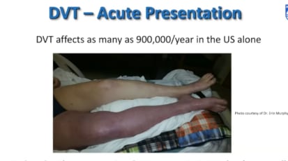
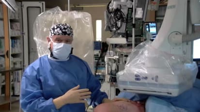
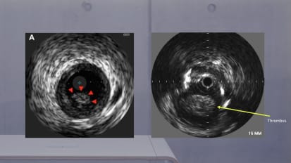
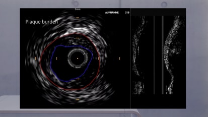
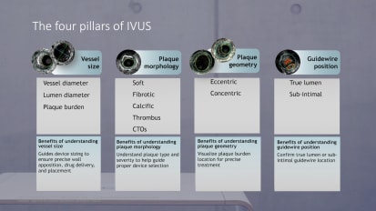
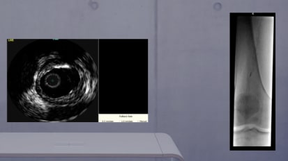
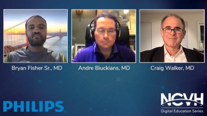
This is an interactive webinar focused on the multi-specialty approach to the treatment of patients with venous disease. This will include didactic discussions and case reviews. The webinar will feature dialog on the overall evaluation of the vein patient, how to treat patients with both superficial and deep disease and non-thrombotic and post-thrombotic lesions.
I have the pleasure of introducing the faculty tonight we have dr steve Elias from Englewood Hospital in Englewood New Jersey. He is a vascular surgeon and is the director of center of vein disease at Englewood Hospital. He is also the founder and course director for expert venus management. Dr Elias is the medical director for a vein magazine and the medical editor of the american venus forums newsletter, vein specialist Dr Anna Safadi is from ST mary's Medical Center and Hobart indiana. He is an interventional cardiologist who has authored several articles on pe his practice has grown tremendously in the area of deep venous disease and particularly in the way of venus compression syndrome and DVT intervention. Now I'm going to hand it over to the faculty and let them say a few words and we'll get started thank you mm. Okay um welcome everybody. We hope that you know you guys enjoy what we're doing here. But more importantly we want to let you know as Nicole already said that what we're really looking for is your feedback your questions. We have a few questions for you, not not in a test form but a few questions to get a better idea about who we're talking to and what specialties. So if you all can vote, Nicole just put up the quick poll as it says, just check off whatever your specialty is. So while they're doing that, do you both want to tell um the audience maybe why you are so passionate about treating deep venous disease and maybe how you both got started? Okay. I just wanted to go first. Yeah, I would be happy to comment on that first and foremost. It's a privilege to be part of this, especially with such an uh an esteemed colleague dr steve Elias. Um you know, it's very interesting and as a segway to the initial question is what is your specialty? I think this is really um uh a key concept in making um physicians and healthcare workers understand that venus disease uh an intervention is something that can really be tackled by uh a wide variety of different specialists. I mean myself, I'm an interventional cardiologist and we were talking about this Just before we started, I would never, in my mind have predicted that, you know, it's about 10 years after fellowship, I would be practicing 60 of my interventional practice venus intervention. Uh and um you know, with that said that one of the big reasons that really compelled me to kind of Get into this field was uh several things number one, I unlike other um arenas or other um parts of different uh interventional type of procedures um that had maybe a saturation or a wide variety of people doing it and it was relatively tough to really grow. I found that venus diseases almost untouched, uh it was, and this is, you know, initially, and even till this day, um uh the amount of venus disease available for people to really be involved in this tremendous and second to that, I found that the the gratification in approaching these patients and taking, for example, whether it's a patient who has severe post traumatic syndrome from a deep venus, uh you know, thrombin arctic event, or taking a younger female pelvic pain. Um and and, you know, intervening on these people, uh I joke around and say this in different lectures that I've given, I get gifts from these patients. Uh I do patients acute am I in the middle of the night, and technically, you kind of saved their life and they barely look at you the next day. And so it was just like, it was amazing to me when I really saw how, um, you know, how it changed the life of these patients and that to me with the combination of the amount of volume and potential, uh really was the key, the trigger that really lit um, kind of the passion under underneath to really get into this and, and kind of, as they say the rest was history and ever since then it's really grown tremendously. So, you know, we hope hopefully today to highlight, you know, wide variety of different topics, but really, you know, depending on wherever you're at, whether you're a beginner and just want to get into it or You're an advanced operator highlight I things that are important things. I wish I had somebody that could have told me 10 years ago in a didactic fashion that, Oh, you know, you should probably do things like this and do things like that and avoid this and avoid that. Know a lot of things that I learned was through colleagues of mine or even unfortunately through things that I realized as I was doing it, I realized retrospectively there's a better way to do it. So, you know, we hope to share kind of our experience and um, you know, steve I'm sure we'll highlight so many very important things that, uh, you know, um, in his talks initially, and hopefully I'll be able to provide some insight as well. And we can really make this a a useful night for everybody. Okay, good. Nicole. Do we have? Speaking of it? Let's see what you have answers to the questions. Sure. Yes. Can we have the pool results, please? Okay, so let's take it one at a time here. So we have most people are others. All right. We have uh it's an interventional cardiologist. An issue could not vote. So that means there's one other interventional cardiology. Yeah. That's right. That wasn't me. Okay. There's no vascular surgeons. Um Okay. And let's see what we say here. In terms of Most people are less than five years And then some more than 20 years. Okay, Very interesting. And yes or you as we have it on this answer. Only bad quarter treat superficial disease and maybe a little bit more treat deep disease. So Okay, that's that's good. Most are beginners. Alright. That helps us out some consider themselves advanced. So we'll try and tailor what we say based upon what you guys have answered here. Most of you obviously have all these things available to you and most of you have used these technologies as well. And most of you have attended a course in the last 12 months honest. You have any comments on any of this? No, I mean as you mentioned, I think um uh the fact that it seems like the majority of the patients are more kind of the begin, although we do have some towards the advanced level, so you know, at least in my part I'll try to, you know, um pose a commentary across the spectrum and then hopefully the most important thing I think is really to open it up to questions, especially since we do have a, you know, a bit of a variety in terms of skill set so we can try to provide answers and um kind of guidance to the spectrum of attendees. So I look forward to being able to, you know, um, to do this together. So I want to give you guys a kind of the big picture, some of you, many of you are beginners and and um even though we're talking about deep venous disease, I just want to give you an overall view about how you need to look at. I think every vein patient that comes into your office setting or or clinic. Um so that's what this first talk is about and then the subsequent talks going to hone down a little bit more on Deep Disease. These are my disclosures. What I want you to keep in mind as we talked through everything here tonight is the treachery of images. This is a painting obviously by Margaret that says this is not a pipe and it's not, it's obviously a picture of a pipe. So in venus disease in general we always need images, but it is rare that we only treat the image As opposed to say arterial disease. Somebody has a a symptomatic, 95 carotid stenosis. We know we have data that if you don't do this, something bad is gonna happen or if they have a seven centimeter aortic aneurysm, that's not symptomatic. So you are treating the image in vain disease, You're almost not treating the image as your primary reason. And that's what I want you to really take away from this. It's really how you look at it, the vein disease and how you look at the patient. So you can look at things from different ways. This painting, you can look at, you know, look at the distance first. You can look near first, but it's you need to think about what perspective do you need to look at that particular patient from? Our main goal of course, is to help patients with quality of life. Many patients are as upset as uh the painting here by monks uh that you know, it affects their quality of life. And what honest was saying, these people are very, very happy when you help to improve their quality of life by something that you do for them. Um and you always need to ask yourself the question, how does it affect the patient that you're listening to? Not? How would it affect you? I mean, we'll see people with huge varicose veins on their legs and they just want to get it checked out. Um They have no symptoms, it doesn't affect the quality of life. You may think, oh my God, if I had these veins, I wouldn't be walking around. So we need to look at it from the patient's viewpoint. And many patients, certainly advanced disease or post antibiotics are like these people in the van Gogh's the potato leaders. They're unhappy if you have an ulcer, you're not looking like you're happy if your leg is heavy and you can't walk around, you're just not a happy person. So we already spoke a little bit about venus versus arterial disease and on in arterial disease most of the time we are telling the patient what you need. I gave you some examples already, but somebody has a gangrenous toe and if you don't do something, you know, things are going to get worse and worse, they may lose their legs In arterial disease. We may ask the clot begins, how much does this affect your life? And that's only about five of the people that we see or treat. I don't treat any arterial disease and vascular surgeon, I haven't done an arterial case in 20 years. But uh the questions to ask are the same. Whether I've treated him 20 years ago or not in venus. It's almost the exact opposite. You got to think about estimation. What do they want to accomplish? What's bothering them? Maybe we do have some good data with ulcers or post robotics or DVT that if we don't treat this DVt, they have a high chance of developing a post traumatic syndrome or ulcers. We have data that if we take care of the deep disease or we take care of the superficial disease when help the ulcer to heal and prevent recurrence. So maybe we're going to tell them what they need. But most of the time we don't. And again, that's the theme that we're going through this year. So I always say you have to be a man or a person with a plan. You need to, in your mind both from history and from physical work, from the diaphragm down to the ankle. When you're going through asking them questions and we're going through examining them and you can quickly do this uh, in most patients is, and that being, you know, an extensive uh questioning, questioning or exam and I'm gonna take you through this. But think of that, that image from diaphragm to ankle, you gotta start from Vienna Kaveh down to the smallest veins sitting around the ankle where the patient may have an ulcer for instance. Um, So by the primary history, uh, you're going to ask, this is what you may ask them any previous DVT. Where was that DVT? Um many times they were told it was just in my leg, but in fact no one ever looked higher up. So someone has history of DVT, You're already thinking I may have to look a little bit higher as well and then superficial disease below the inguinal ligament. I want to ask what we call a hasty or Heaviness, aching, swelling, thrombin throbbing and itching. So you're asking about those symptoms, have had any previous vein procedures and have they had any ulcers. So again, you get some idea where the problem might be. But you want to ask them about these things. Primary exam from diaphragm to ankle. I think in almost all patients, certainly those who have previous DVT or you think have post traumatic disease, you need to look at the abdominal wall. They have any abdominal wall, viruses that are acting as collaterals esc esc. And look for any viruses in the groin area, The labial region, the pharyngeal region. Look at their ankles, skin changes, previous ulcers. Do they have uh you know, a trophy blanche, which is shows that they have advanced disease. So you need to go diaphragm to ankle varicose veins. Where are they located? Are they in the typical medial calf, a medial thigh or they located in other areas? And I'm going to go through that. So that's your initial primary exam. And honest, if you want to jump in at any time, you know, and anything feel free to do it. Um how about so now you've done your exam and you see veins like this, This is not the usual location of varicose cities on the inner thigh or anterior thigh and they look like they're going up into the England region. Are they origin may be higher up. So when you see something like this, or dark Aziz on the posterior thigh, that going to the glue you fold? You may want to think about asking some other questions because now, maybe you think this is even though they came in with varicose veins in their leg and some leg symptoms. You want to ask about other pelvic symptoms, do they have significant pressure as they're standing during, during the day, as the day goes on? How about when, when they have their menses is they're paying kind of worse than most of their friends or they think it's kind of added out of the ordinary. Do they feel pressure on the bladder as the day goes on? This is all asking about varicoceles within the pelvic area. Do they have post quote, Okay. Not pain during intercourse. This peru nia is something usually a something within the the ovaries, the uterus, but post coital pain. And when you when when you patient has this and you ask them, their eyes light up, you say not when you're having intercourse, but 15, 20 minutes after intercourse, you have significant discomfort in your pelvic area. And they said, Oh my God, I thought I was crazy. Yes. And that's because all the blood was brought into that area when you're having intercourse. And now, if you can get out because you have venus disease patients have this post quota pain. When asked them in terms of leg symptoms. If they've had previous DVT, ask about venus qualification and then you need to ask them if they have a combination of symptoms. What bothers you more your leg symptoms or your pelvic simple anything on this out? Because I know you I would say another thing that I've noticed uh in terms of physical exam, in in terms of taking a history, uh, that if you actually pinpoint to the patient because sometimes they take it for granted or don't don't really think about it as something I realized when you ask them, have you noticed a difference and the size of one leg over the other, particularly the left, bigger than the right? Um, a lot of patients that have come to me and obviously this is a select group of patients will ultimately end up seeing me. I find that they say, yeah, you know what I actually, it's funny you mentioned that, you know, my left leg is for some reason it's always been bigger than my right now. It could be the opposite as well, but more commonly the left side. And that really to me as you, uh, you know, no, can be a very, uh, a very important sign that there's something going on. Proxima proximal uh, in the deep system. And that's something in addition to everything you mentioned. I think if people can hone in on that, I think it it provides a lot of utility and just in terms of the simple physical and history uh in patients. Yeah, that's a good point. So the other thing you need to remember when you're taking care of aging patients is that vein disease really is an incurable disease and well, it may not be too good for patients, is kind of good for us. If we're treating vein diseases, nobody's going to come up with a cure for vein disease. Certainly not in my life to have more than a lot of people here, but they're just not. It's not a big priority to cure vein disease. So you need to understand what your goals are in treating these patients and let the patients know what they can expect going forward into the future. Mhm. So as I said, you need to define the path of physiology. Is it into the pelvis, Is it in the leg or both? And again, that all goes down to going from diaphragm to ankle. Pelvic symptoms can be caused by either compression of the veins or reflux. Uh leg symptoms can be caused either from the pelvic area escaping down to the legs or just disease below the inguinal ligament. Whether it's reflux the superficial system and varicose cities or some type of obstruction. Post robotic disease in the veins of the leg, the formal vein portal vein, there can also be reflux of the internal iliac going or ovarian veins, thermal veins. Patil saxophonist, varicose cities, compression usually is of the iliac and or the renal vein post robotic can be either in the L. Yaks, in the Vinick Eva were in for inguinal in the the deep veins there. So you should get some idea about where the problem is. As you speak to the patients, there's a huge differential diagnosis. We're not going to go through all these, but these are the things to keep in mind. And this is why people may come to you with veins that they see, especially in the leg or even in the health care. But veins on the leg symptoms on the leg, but and they're trying to make a plus B equals C. It doesn't always equal that. Certainly not in leg symptoms. You can see this is just a little list of what's in the leg. So I cannot tell you how many times I've seen agents who come to me with veins on their legs, they said they've had a procedure, it didn't help. And you ask them what the symptoms are. And they'll tell you, well, I have a pain, it kind of starts in my buttock and it goes down the lateral aspect of my leg. Well, of course, it's not venus that's, you know, say attica or they have Leg swelling and they wind up with two arthritic knees that need a replacement. So, but they have some veins on their legs. So again, you need to try and put it all together when, when you're talking with them. So, your first question, you need to ask yourself after you've done everything history and physical and your secondary, what I call history. Once you've seen where the veins are located, if they're abnormal areas, you first as they are the symptoms or science venus. And if it's no, you're done. If you say yes, then you need to determine did it affect the patient's quality of life? You asked them all these questions. If it's no, then you're done. Because we have almost zero data that says, if someone has vein disease, but no symptoms don't care how it looks, never had a complication that you need to fix it to prevent something in the future. We have data with other diseases. Of course, we have data with material disease. We have data with uh colon polyp etcetera, etcetera, but not in venus disease. And that's why I want to highlight. We want you to be treating the right type of patient, not the image. It needs to affect the patient's quality of life. So if it does affect the quality of life, well then you're going to go beyond the history and physical. You have to decide what patients not to treat. Here's my list. Obviously they don't have symptoms are in vain and they don't have any vein disease. When you investigate them, the symptoms are in venus, but they do have some vein disease, like the patient was psychotic, the symptoms of venus. But you try your hardest approved by imaging and stuff. They have a disease and they don't um and they have symptoms. They have vein disease. Again, it doesn't affect the quality of life. You don't need to treat. So how about what do you need next? Once you decide, yes, it affects the quality of life. Yes, it's venus. What do you need? The question is, who doesn't need an ultrasound? Almost every single person needs an ultrasound, maybe the purely cosmetic, tiny spider vein person. But after that, almost every patient, you're going to see that once you determine the symptoms of venus is an ultrasound, um and then you determine, am I just going to go from the groin down? But I need to look up as well. And that's going to be determined by their symptoms and what you found on exam. And then rarely we'll go to more actual imaging to look at pelvic disease and we're gonna talk a little bit later on your seat, patients who do need videography and intravascular ultrasound. Um, so now the patients to treat they have a venus cause it's from the pelvis or their legs or both. It affects their quality of life. The image correlates with what you have seen on them and gotten from them for their history. Again, we don't treat the image. The image has got to corroborate when you've decided it's venus disease and you set expectations for yourself and the patient honestly have anything up to this point. Yeah, I want to highlight that one. I think that's extremely important. Important to drive home the concept of deciding on one to treat what to treat, how to treat and what imaging modalities. Uh, and I think like a case example is if somebody comes with leg oedema uh maybe a little bit of discomfort for maybe like a kind of a C. Two to C. P. Three patients. Um You know, my threshold to be very aggressive and do very invasive things is very different than someone who has an active sihp six venus, also both in terms of deciding what to do with the patient in terms of invasive evaluation, whether it's something like the C. T. V. In a grammar or or actually take them to the Cath lab for Ivan's venogram. Uh But in addition to that, if I take him to the Cath lab and do um imaging both venogram and intravascular ultrasound, the threshold to um decide on when to treat how much areas stenosis or compression you have is very variable. And this is not the case with other disease process, other disease processes. We generally have kind of you know, wherever that is in the coronary bed and the karate kid in the iliac. We have general guidelines that say, well if you have, you know, if you have 70% more crowds cyanosis or 80% that's you know, generally considered severe, you can do a B C. D venus work, that's not how it works. There is first of all that there is really no widely accepted guidelines in many different areas, but specifically when you decide to treat and how you decide to treat is extremely variable. And I think um these comments about you know choosing based upon the patient's symptoms and also you know, be kind of what they bring to the table and and the severity of their disease process is extremely important. And making clinical decisions on these patients Because I I've had personal anecdotal experiences of patients, for example, seats six and they had maybe 40-50 compression of their vain. And we'll show examples of that later And that their operator left it alone because it, you know, wasn't 50 or more if you're using that as the uh just kind of general guidelines. And they came to see me. They had, you know, continued venus ulceration, not healing. And I ended up doing them and stenting there 45 compression because I felt that they were in a different situation than, you know, somebody who just has like pain for example. And they ended up doing very well and their symptoms improved in their ulcer um, uh, heals quickly. And so I think the concept steve is highlighting. I think it's something that is very different than other processes that all of us, as physicians have kind of been taught and we have to really apply to. You know, take all the other hats off and put the venus hat on because it's a different hat. And I think if you can go away with that concept really understood well, I think that is a huge benefit and I really like the way he laid it out. I think it's extremely important to understand that. All right, thank you. All right, So let's go through that. Good. I'm glad you highlight that now. So, these are the questions you ask yourself and and honest just said that. Is it super. Does he in for single disease? Super England or in for England or both? What is the minimum you need to do to help the patient and then think yourself. What is the maximum each occasion has, what I have called a hypertensive threshold. That gives them symptoms or ulceration or whatever it's different for every single patient. So you need to figure out where this patient sits on that hypertensive threshold uh line. And what is the minimum you need to do? And what is the most you need to do? And you need to remember? You don't need to do everything at once. As long as you have, as I said, a man be a man with a plan. Um, so there's black and white and there's gray areas. If someone people ask, well, what do I do if they have, you know, pelvic legs? And what if they only have pelvic, then you're going to take care of the symptoms, then you're going to take care of the pelvic disease. If they have leg symptoms and they only have in for England disease is going to take care of that? or what if they have combined symptoms, you need to ask them what's worse for them? Did you like symptoms bother you more or your pelvic symptoms bother you more and that's where you, what you attack first. If they say it's both the same, here's where imaging may help you if you can determine which imaging you feel is worse, so to speak, and that's still a great area. But usually you can narrow yourself down pretty much to uh, knowing what to do if you ask the right questions and then let the imaging kind of push you in one way or the other if you're undecided. Um, if someone has like symptoms, where's the pathology in the pelvis or the legs are combined? If it's within the pelvis, um, you may treat the pelvis. If you see within the leg, you may treat the legs. And again, if it's combined, you're going to treat which is worsened. But usually most of us, if it's combined disease causing like symptoms, we may treat the uh leg disease before we'll treat the pelvic disease. But again, as an, it's already said, there is no clear rules on this. More importantly, you need to go slowly. You don't need to treat everything at once, you go where you feel, you're going to give your biggest bang for your buck and improve their quality of life. It just has a little aside, anything you do to a patient regarding venous disease, You need to tell them it takes at least six weeks Up to 3-4 months to see the full effect because if you're you're someone with an incompetent, great staff in this vein and varicose city, say, and you just treat the sophos, which many people are now doing. But that's not my point. My point is you oblate the cevennes that's causing the symptoms and the visible varicoceles. But those veins have been dilated for so long, it takes them at least six weeks up to a few months to shrink down to improve symptoms. And Kaz Missus. You stent somebody, sometimes people tell me two weeks later, a week later, oh, I feel so much better. But still you need to tell them it's going to take at least six weeks up 3-4 months. It happens sooner fine. But you said the approximate disease, all the veins below have been dilated for so long. It takes time. It's not like an ischemic limb. You re vasculitis and boom, it picks up and you feel the pulse that everything is wonderful. Get put on your venus. Hat is honest. Um, so in conclusion, who needs treatment? Well if you're treating the right patient, you're almost always gonna treat them. Almost never going to treat the image alone. And you're going to treat both the image and the patient when the image and the patient all fit together. So remember the treachery of images. This is my son. A few years ago we were in Greece at a meeting and here's the gym. Well here's the gym. So the image of a gym that I had when I opened that door was not what I saw once I did I did open it. So have a plan B. The right doctor treated the right patient for the right reasons. You'll be happy, as Ana said already because your patients are going to be happy. That's it for now, honestly. To addition and I think we have one question um from what I understand, so I think we'll go ahead and take that Nicole if you can help us with that. Yeah, sure. So there's a question and again, I'm not sure how well the two of you from your viewpoint. Because it's a question about the difference between mobiles and hospitals with choice of access points for deep venous disease and both of you guys are hospital based. So what do you mean? Choice of access point access points on the leg where you would access? Yeah, believing that's where this question is being directed. And do you think it matters? Yeah, I think I I well, I mean, as you mentioned, I'm, you know, I'm uh employed and work in a hospital type of setting. My probably my guess is this question is going to the point that, um as we're gonna get to, when we talk about cases, you can approach venus, um, interventions and a wide variety of access points. Um um whether we're talking about the robotic or non traumatic. Uh so for example, you can go into the common from Mulvane, the proximal femoral vein, the great staff, and to stay in the pop little vein, the posterior tibial vein. And so I guess the question is probably trying to get to is there a preferable approach if you have like an outpatient lab maybe in terms of allowing the patient to be discharged quickly? Um You know, obviously the smaller the vein access uh the easier the recovery is from it, yet the flip side to that is you have more limitations in terms of what you can do to that patient. So um I really think firstly to answer that it depends on what you're treating um uh you know, uh what level and what type of technology you have available to you because that's gonna really that for at least for me, and we'll talk about this shortly when we get to the cases, that's how, you know, I come to the decision of, you know, um which access point I want to use. So I'm not unless the physician can specify exactly what he or she means by that, I'm assuming they're trying to figure out is there in access um point that will be kind of the easiest allowed patients to quickly recover and quickly get out um from an outpatient facility. I think maybe maybe there might um looking at maybe do you both have a preferred access access site or even is that even a thing as having a preferred access site? What you're doing? I don't think it is preferred. First of all, it's what the patient gives you. Um I mean, you know I happen to like the great staff in this vein as long as you can use that because you can you know, access into the sadness and not have your sheath even go into the common thermo. So you can see disease from common thermo all the way up. Federal vein is very nice. Also my least favorite access is actually pop little. It may be easiest, but I think for the patient certainly if you and and every almost everybody I I do does not stay overnight unless they need for some other reason. And I also work in a hospital setting. So um papa tells the easiest and maybe the most comfortable for people. But I think you should start thinking about sadness or femoral vein. And um that I mean that's kind of how I feel. I don't know what Yeah. Yeah. So again it depends on what I'm treating, but if we're talking about like venus compression on prom biotic, I'm a fan of proximal femoral vein. Uh and um the only time I keep patients overnight and I was going to get to this, because when you were talking about, you know how to approach the patient and what the patient should expect and what the time frame of recovery is a big issue that I spend talking to patients before doing the procedure is the explanation. And and the clear understanding presented to the patient, that if you have a compression syndrome in the iliac bed and we spent it, you will have pain. And um these patients, if you don't explain that to them uh and they don't know what to expect, they will be horrified. Uh And so I keep patients a lot of times just for pain control. Um uh though not everybody if they're not having pain within the first few hours and they're not gonna generally have pain, but I've had my share of patients that had a lot of pain and tell them what pain you're talking about. Yeah. So, you know, obviously um when you're stenting the iliac venus system, you have nerve plucks i that are there and you're you're you're you're you're also stretching a vein that's completely compressed shot and almost ripping it back open. Uh and you're pushing against, you know, uh the back structure as well. And so the combination of that with the pressure with the nerves. Um These patients have, and I tell them, and I explained to them before, just know whether we're doing pre dilation with the balloon or actually deploying the stunt. Um um and I found that if you explain it to them and really kind of guide them, they tolerate it and they do very well but if you don't do that um they're horrified a lot of them. And so um that's why one of the also potential issues of um you know, trying to discharge the patient the same day. Sometimes it's paying control and we can talk about more more more about that when we go over the cases. But um I agree with, you know, steve, we try to get them out the same day if we can, but there are issues and if you're going to get into venus work you have to, you will come to terms with this. I mean there this issue is real and and it's something that um you know, as we mentioned, you really just need to educate the patient on yeah. Any other questions that call or we're going to go on. Well let's go on out. There are a couple, but let's go on for now and then we'll get to those. Okay, so here's the case of what would you do? And we'd like to hear from you guys if we can get, what would you do? Um Okay, here's I've been compensated. You see this here is my disclosures. We saw that already. All right, so keep in mind what I The talk I just gave you before this. So here's a 23 year old female triathlete. She's very crosses of Kiev medial need for the last five years or so. This swelling, this robbing, there's a king citizens are definitely worse at the end of the day and also during, during menses, she says her legs bother her more um compression therapy, compression stockings does help. There's no history of DvT or S. V. T. Honest or anybody else in the audience have any other questions on the history. I'm going to get into physical and stuff later on. Um I would ask a patient like this, you know, and this goes to the commentary you presented, you know, is where what part of her pain is more severe. Is it the pelvic pain aspect of it or is it some more a little bit of the kind of superficial system aspect of it uh to kind of decide, you know, how to approach the patient first or where do you focus in on first? Because that's yes, she doesn't have any public symptoms. This swelling, probably an aching. That's worth the end of day, during menses is her lower extremities. Okay, so she has no pelvic pain during meant, no. Okay that's that's that's what I was trying to do. So on exams just got good pulses and you always need to take pulses even if they're young. Um She has varicose veins and left medial calf and around the knee. There's 6-7 mm, mild calf swelling. Just have any skin changes or ulcers, Basically AC two C 2 1⁄2.. Any other questions on physical example, does not have any abdominal wall viruses. Duplex showed no acute or chronic changes. Uh, DVT or post previous dVt, there was an incompetent common formal vein with some reflux About 1.8 seconds. The federal vein and the property of a more confident Great staff in his vein nine. the calf measures 7.5 millimeters and the reflux times greater than three seconds. Why do you feel like there's a reflux potentially in the common femoral vein with the history like this? Right. So what I was, what I was going to say is there was kind of a maybe a little bit of not fully physic flow in the common formal vein also. Um Why do I think so? Well, maybe we better see why? Yeah, Well, I just I'm highlighting because I think it's important that you mentioned the lack of the basic flow just to guide people and understanding in terms of central or more proximal type of disease processes that may be playing a role here. Right? So we're gonna ask, ask all of you. And if you if you don't vote for any of this, then we'll assume that you think we need something else. So most people want to look somewhere else. Most people just want them to have compression. So we have yet interesting. Would you just tell her to keep an eye on that? Isn't that would not. I mean, that is interesting that that was the majority of response. I mean from what I see it's a younger lady very symptomatic with significant reflux and the DSV. So you know uh probably failed um or attempted conservative therapy and failed it. Uh So I would think that you know something uh to be offered her like you know gSP ablation or excision, you know if if needed as well. The very cuisine would be something maybe as the initial start and then potentially based on some of the ultrasound imaging you show it. I think you would also have to really keep in mind the issue of ruling out some type of venus compression uh disease process as well as this patient. All right, so we thought of that. I thought because first of all vascular text are very savvy in terms of you know, it's not a normal flow in the common from, well they may look above. And also this person remembers she's 23 and she developed varicose is at the age of 18 which is relatively unusual. So you may want to think is there something else going on? And she has a pretty big staff in this vein for being a young woman. So is there some something happening higher up? So we decided to do a venogram and there appeared to be perhaps some narrowing here. Can we run this again? Yes, we can. An external iliac vein. You can see the veins below are a little more dilated. Then then what's above? And when? At the time of the venogram of course we did a intravascular ultrasound. So we're starting up here and I did this kind of slow. So we're up here in the vina cave and we're coming down and you can see here we're now this is the aorta right? Coming here pressing down and that goes away and now we have right common iliac artery were in the common iliac vein on the left and it was going slowly here and now we come and we see the internal and external arteries and we're coming down the leg and see some compression here as you go. Will further down. We see a little bit more and we'll come below the area now in distal external common federal vein. Yeah. Okay. So first of all, honestly you think that this is a real finding? Yeah, absolutely. It's not a trick. You know it's not a trick case. Okay, so we have a real finding. Okay now can you give them a choice again? You can. So we'd like you all to vote as to what you might do now. Alright, how about those results? 88 are going to angioplasty and stent? Okay, honest. What would you do? Uh I think based on that I've issue um which I think is also important to highlight how even on the venogram we were talking about there was some compression. You know that we saw an external like me. But what I what I want people to take away the difference of the severity that you saw an ivy is compared to the venogram. I mean you saw that distal external iliac vein um mid externally. Like it was almost like a split. It was a slip like type of lesion which is you know very significant and makes uh uh definitely makes sense. I think personally given that in those findings I would first start with treating the proximal and seeing how the patient did it decompress the leg and reduce the reflux and because especially she's so young kind of then so be it. As you mentioned, give it give it time. This is not going to get better in a day or two. Give it a couple of months, see how she does. And you can always go back and if you have to do adjunctive ablation or other therapies to proficiently then you know then so be it. I guess I maybe I may be wrong. Okay. The next technical like secrets in but we'll move along because the next case is very, someone can do it really quick. So remember I said, what does the patient want to accomplish? She does not have significant I don't think obstructive type of symptoms as opposed to reflux in the varicoceles and the great sadness. It's only leg symptoms and there is some swelling, but it's worse at the end of the day. It's not in the morning time. She has some throbbing and aching, but no venus qualification. She is a triathlete. So she's not getting significant qualification when she's stressing herself. So I She's 23 years old, I think, A long, long time before I'm putting in a stent in a 23 year old personally. So we, we talked about this what affected quality of life. Her leg symptoms affected the quality of life. I thought that staff, this was big enough. Um, and again, as I said, you don't need to do everything at once. You don't burn bridges placing the stent in a 23 year old. In my mind is the first line of defense, so to speak. I didn't feel so comfortable that she was a 56 year old. Yeah, no issues here. Um, so here's what I did. I did an ablation of this happiness game. four months later, most of the varicose cities uh got small enough that I could do ultrasound guided scleral. Her symptoms improved. And she really had no uh no pain at all. That's what I did here now. Maybe it's crazy. But I explained everything to her and her parents and I said, here's how we're gonna start because I think your symptoms that bother you may be coming more from your great staff anus rather than you are what the image showed. And again, that may have been the wrong thing. But in this patient it wasn't helping her now. She may be something further down the road as this make us get worse. But for now I just didn't think the first thing to start with was to put a stent in, in the patient her age, there's no right or wrong. But in this patient worked out now. How about this patient, which is going to sound very familiar to you? 23 year old female triathlete, Varicose veins are left calf and media only for five years swelling, dropping bursting pain after her training worse at the end of the day, she has not used compression. But then after I asked her to use compression, she actually said it made it worse as the day went on. Any questions honest? No, no, no, not yet. Okay. Same exam as the other patient who was the 23 year old triathlete. But here her calf was somewhat tense when she was standing for a while and there was no skin changes or ulcers. So similar findings, exact same findings as we had in the last patient Nicole. I'm going to skip the questions. So here here are our choices. But here we did some more work up which, believe it or not, is the same exact findings as we saw the last time. Exactly the same findings. This is the same exact Davis. Now, can you put that up again? Nicole? Yes, we can. So you mentioned on history this, this lady had more uh more symptoms with exercise. Is that correct? Absolutely much worse. She had trouble training. Pain at the end, bursting pain in her calf as if it's going to, you know, quote explode. Okay, We should get the results, you get honest and I are laughing. Okay? We got to browbeat you guys into into this. Okay. So lets people want to do a stent in this person. All right. Maybe I was wrong again here, but here because her symptoms, I thought were more from her approximate disease. She had venus Claude occasion. She had a tense care when she's just standing. She could not tolerate compression stockings because her problem was outflow obstruction, not reflux. If somebody has reflux stockings in general, make a difference in symptoms. If they have outflow obstruction many times, they'll even say it is worse because now you're squeezing against an outflow obstruction. So here, after blaming, this is actually what I really did on this patient. The first one was made up to make the point of what I was talking to you about about. You need to see what the patient wants to accomplish. So, no. In this patient I really did because her symptoms really were uh Related to the proximal disease. But I'll tell you, it took me three times over the course of six months talking to her and her family to understand what she might be getting herself into because we don't have data placing stents for the next 50 years. And the cevennes actually became competent four months later, I'm sorry. Savviness actually became competent. My phone. I mean I I want to highlight the importance of the comment you made. Um you know, I have a relatively large patient population of young females because I deal with, you know, my obi gynecology, gynecologic colleagues and me a lot of a young patients who have unexplained pelvic pain. So you really need to document and discuss uh the issue because inevitably if this can of worm is is open in the sense of you start um evaluating these patients, you're gonna find venus compression and a lot of times that is going to be the cause of their pain. But you need to document and explain to them because it is true. We don't have long term pay agency data. There are uh in terms of um uh iliac venus denting in 23 25 30 year old patients. We don't we have some anecdotal data, observational data on pregnancy instant. I have these discussions and I document, you know what about anti platelet therapy, antique regulation. You know, all these things have never been delineated in any large trial. It's all expert opinion, quote unquote people do what they think is right. People extrapolate from the arterial bed to the venus bad. Uh and these things and these are not as Steve mentioned is not a 67 year old patients. These are 2030 year old patients I think not offering the therapy because of their young age is problematic, but because a lot of times is the cause. But I think at the same time you really need to document and and have discussions with the parents with with obviously the patient and really make sure you know these things are well clarified uh you know for the patients sake and for your sake for that matter right. The point of both of these cases is to make you understand. You need to start from what the patient wants to accomplish and not the image and what symptoms are being caused by what uh first case I thought it was mostly in for England problems. She didn't have a bursting painted another's this case here. Yes, I thought it was approximately this is just a stent. You see whether two arteries were compressing the vein before and and and she did well. But again, food for thought how you approach the patient and you just need to ask the right questions and ask yourself the right questions as well. So there was a good question that came in about like symptoms with negative venus duplex. Can you comment on C. T. V. Slash MRV verse venogram? As next imaging modality. Mhm. For leg symptoms, if they negative with a negative venous duplex blake symptoms that you they saying that they think it may come from higher up. Yeah, they're just saying that the patient does have like symptoms they've gotten, they've gotten the venus duplex which has been negative. Can you comment on ctv verse MRV first venogram as next imaging modality. First dot com. And then so first of all, um if we're thinking that this is a problem higher up and the symptoms are venus. Remember not psychotic or something like that in the leg. First of all, we get a trans abdominal ultrasound which most good technologies, once they know what they're looking at, you can see if there is a compression or narrowing if they don't and I personally if they don't and I think there's symptoms really are venus and they really have a chance of having something higher up. I will do a diagnostic Ivascyn venogram rather than going to C. T. O. R. M. R. The only reason I ever go to sea to um our first is if I think there's other pathology in the pelvis, tumor fibroid what everyone do. I think it's venus compression. I will go to Iverson venogram diagnostic not with intent to treat because if you get a CT venogram or MRV and it shows some compression you still need to do. And I have some venogram to document how good or bad is that compression? If it doesn't show compression and you still feel the symptoms of venus. You need to do a venogram and I've is to document if it's normal or abnormal. And the calculation venogram and I've this they wind up with a band aid Wherever you did your access and they walk they walk home or whatever they need to do. So it will give you 100 of diagnosis without radiating them or whatever else. That's it's a little bit aggressive but that's the way I reason it. What do you think an issue? 100 agree. So first of all the majority of these patients when they finally end up seeing us, people who deal with this, they've been through pain and symptoms and work up for years and years. I've had some patients who have had 15 20 gynecologic procedures continue to have for example public things. So knowing that there's a test modality like C. T. V. Program that has sensitivity specificity is in the 60% range. To me it just does not cut it because as steve mentioned if you do it and it it confirms it then you still need to move to Vienna Graham and I've if you don't do it or if you do it in it and it's supposedly um rules it out it doesn't rule anything out 40 45 50%. Sometimes patients may have it. So I personally don't. I mean I I talked to the patients explain them the options and whatnot. But I'm a big proponent of, they've come to me, they reached me especially if they're more advanced disease. Um You gotta go where the money is that you've got to answer the question for the patient. Uh Number one the only time I incorporate personally see tv anagram is if there's an issue of um you know access points and chronic total inclusions and you need to kind of figure out well you know which access point should I use to help get through this. And it's a more complex type of case. N. C. T. V. Program can be helpful for you. But just general standard work up I've I used to do more of them. I do way less now. Um For the reasons that steven and I mentioned it, I'm going to focus more on the aspect of deep venus de venus graham biotic type of interventions in cases A little bit different than the non traumatic that Steve showed. And so what I hope to do is kind of walk you through several cases uh and uh and also again as we do it um if there's any question throughout please let us know and we'll be happy to to address them. Uh so in starting um this first case is a 30 year old lady who came to see me with several year history of left side of pelvic pain. In addition to that, she also obtained um and the left leg and swelling uh really predominantly isolated to the left leg. She said, you know, when she would stand for long periods of time, that's when her symptoms would get worse. Uh So, you know, when I first saw her, I said, well, as kind of uh steve mentioned, you know, how do we work this up? What should we start with? I always get venus reflux studies. And then I said, let's talk about the results of that. And and then depending on how you're doing, we can and and and where we go from there, we can decide on further work up like venogram and Ivan. So then she actually got lost to follow up. She never followed up and she never got the venus mapping, and I didn't actually really know anything about her there after until she presented um Mhm. About a little over a year later, she came into the hospital which, you know, um significant left sided like pain, swelling, redness. So at that time in the emergency room she had a venus ultrasound done, which showed an extensive dvt uh involving uh again at least in our lab, you know, and as is the case really in most hospitals, I would say in the country, they really, you know, they really just go from the growing down an ultrasound and that has its problems. But as you can see in the ultrasound, she had extensive caught, mainly, it was in the left common informal vein. Uh papa still has some, but it wasn't a clue. Uh So obviously she's a young lady, she's very symptomatic and proximal um dvt. Um And so um kind of before showing these this v anagram maybe I jumped the gun. The question is you know um if you're gonna do a case like this whenever you approach a deep vein case I think the first if you once you've decided you're gonna do it I think that one of the first questions is your access points. And and generally speaking and I think this is a very important concept that I didn't understand as much initially and my um uh experience with D. V. T. Is you really need to go open to open. You really need to find a proximal aspect to wherever you're treating that's relatively open with good flow uh and try to open throughout the course of the rhombus and then an exit or up this whole aspect that's open because if you don't do that you're gonna have a lot of issues of re inclusion. So this latest papa Sylvain, an ultrasound has good flow, a little bit of residual thrombin but definitely easy enough to access through the pop little vein. So as you can see in the picture over here this is the mainly you're seeing the femoral vein but the public will gain is kind of cut off at the bottom and then you have extensive which is actually inclusive in the mid proximal segment with a pretty large collateral coming off this venogram picture here is actually the iliac system from the common from Mulvane up and again what you can see if you have very extensive um uh rhombus nearly exclusive and uh kind of correlates with obviously the findings that we uh that we expected with this patient. So um I'm gonna uh kind of see what you know how steve would approaches or anyone else for that matter based on kind of this. And I will I will definitely um uh say there's many ways you can slice this but I'll just give people just a few seconds to kind of think about it and then I'll kind of walk you through what I did and and and what what um what one should take away from a case like this. Okay so you know I don't know how old this cases or not but I am if you can and there's no metal in the patient which there isn't. I tend to like to start with a non from ballistic device to remove as much stronger us as I can before I begin to put uh some type of Ron politican T. P. A. Or something like that. So in this case I might actually start with like a cloth retriever or penumbra or something like that. That's going to mechanically remove whatever thrombosis I can and then if I need to clean up with something like angie jet I can do that. Yeah that is kind of where I think the algorithm goes in my mind. I start with non throwing politic can and then go to go to a throne politic if I need to clean up or if I need to drip them overnight with echoes or something. But first I start with yeah I I like that algorithm I would say. Um I I have moved more towards that more recently using the Honoree device like you mentioned the clot retriever. Um but this case was done before we had that device so I chose to use equals pharmakom mechanical coming back to me. So we placed a 50 centimeter catheter which is the longest catheter. And the reason it's important to understand catheter length and treatment length because it's important in terms of the access point. Uh and and that's why understanding where you want to access is also important because it provides potential limitations. Um for example if you go posterior tibial vein and you try to access, you know from, from uh you know all the way down above the ankle. Um let's say you have an inclusive pop appeal vein, you're going to be limited potential and sometimes on the size of she usually six french, maybe seven french maximum but usually six french is all you can get in. And yeah that will be enough for like an echoes catheter. But adjunctive therapy may be limited so you have to always keep in mind, you know what what what you're trying to treat and what devices you want to use. So the popular sylvania DVt generally as long as it's not, you know, um heavily involved in exclusive is lens to um many different devices. So as as is shown here, the echoes device was used. So we did catheter directed accelerated license overnight and then you can see here this is the next day so you can see the seminal vein has cleaned up. There's some residual my mild on. But I'm a big proponent of adjunctive therapy. I think that um, uh, people who do don't do adjunctive therapy a lot of times you're gonna, you're gonna cause problems to the patient, particularly when we talk about adjunctive therapy. There's a wide variety of things. You mean balloon maceration or angioplasty is very commonly used, kind of below the groin and then above the groin. We're talking more very aggressive evaluation for compression syndromes. Uh, and by the way, this is a side and I don't have much time to get it. This is one of the problems with the attract trial, which is one of the big DVT trials. Is that adjective therapy was um there was a wide spectrum of variation and um and I think that uh is a big problem with the trial, but that, you know, just to throw that out. So we did balloon maceration angioplasty. Um and and unlike the arterial bed, you put your venus hat on and used bigger balloons here. So, you know, um in the femoral vein, you're using eight and 10 oh balloons in the common femoral vein. You're using 10 and 12 oh balloons very frequently to really aggressively macerate and and get good um angioplasty result. Uh and and so we use, you know, 1080 here and the femoral vein. And we use 12 60 I think in the common formal vein. And then after that we did the anagram. And then we of course focus our attention on the intravascular ultrasound. Now I want to remind you this is the lady that a year and a half ago, um or roughly a year and a half ago had symptoms very worrisome from a southerner. And at the time we talked to her about, you know, things like the instagram. And I've it's and it's interesting, I haven't seen too many of these, but I've seen them where this actually was probably the night is what led to her clock uh potentially. And so uh let's see, we play that I've us again. Um So this is um I was playing again as you can see this is interfering. Okay? But there's the Aorta at 9:00. And you see how it has that compression right at the office team of the left, come in the mail. Uh and then it's kind of slowly going down um uh you know, they're after. And so um the importance of this is really making sure you as we talked about regarding um dVT intervention and adjunctive therapy. The final piece of the puzzle is you've got you have to um open up that compression syndrome that uh that uh potentially was the trigger and also played a big role in this. So when we do that, um there's several things that, you know, we need to talk about. I showed the image, but what I want to show here, which is really important is um um you know, when you're unilaterally accessing, you know, um sometimes you'll have um you'll see the confluence, but sometimes it's not as clear as you want to uh as your, as you um necessarily want and you really need to delineate the confluence and the optimum where that compression happens because that's where the money is in terms of placing the stent. And the nice thing is that the newer night in all steps that we have the venus stents that are available to us. Um generally you unlike the older walls that you can be very exact with them. And so sometimes when I'm not 100% sure I do this up and over trick and I just kind of push it down just to clearly show the bifurcation of the ibc. And I just wanted to show that usually I don't have to do this. Usually I take the tip of the ivies catheter and when I see the confluence, I actually flora save that and then I know exactly where I want to place. You know, the the proximal aspect of the stent. I typically whether you're using beach, even noble, which are the two um, being a stent that we have available presently you can flower the stent just Maybe one cm above where you want slightly bring it down and then once you like where it is, you release the rest of the stent and deploy it and you can be very accurate. Uh and uh and again, that's something that we didn't have with wall step small sense. We would have to hang up into the I. V. C. A couple of centimeters because you wouldn't know necessarily how they would behave. And I think it's something that, um, I personally really really find that the newer generation stents have made a big diff. So we ended up placing a 14 by 1 20 vinovo stead. Uh, and, and, and the patient did well, a couple of things about venus stents um that I think I want to highlight, particularly those who are kind of new to the field. Unlike arterial stenting, you don't do the spot stenting and short stents right at the segment. That is something that you should not try to do. Generally venus stents are long and you want them to be long because you really need a nice anchor uh to be placed in the normal sections. So you go normal now, essentially normal and you have great anchoring and you, the risk of migration uh is very minimal with this type of technique, particularly when you're sizing the stent to the um uh to the appropriate size using IVF. Uh In terms of sizing, another point that I think is important to understand is uh you generally don't want to size to necessarily the priest cyanotic area in the common iliac claim that the mistake that um I see a lot of people potentially doing, I even did it early on. In my experience, you really want to size to the normal segment. So if you have for example, a compress hostel, common iliac vein and then you have a very pristine arctic area just proximal to that. Then right at the distal common iliac vein, external iliac vein. The size of the the vein really comes down and then forms that nice kind of tunnel appearance. Then you really want to size to that area because that's going to tell you what the size, the normal size and you and unlike again the older spent like the wall percent when you're using night and all sent. You really want to size, You know, 1-1 sizing. You want to be accurate. You don't want to oversize like we used to do with wall stenting. Uh and also that will also lessen the issue of back pain. If you sit there and oversized extent aggressively, you potentially may have, you know, a lot of problems with back pain for him, potentially long period of time. Uh So I'm going to so yeah, go ahead steve please a couple of comments. First of all, I think there's a great comments. Also the reason you want this to be longer. You want to set it in nicely, but you do not want to end the stent deep in the pelvis. We're looking here at an ap view of the stent that it looks like it's a nice straight step. But if you look from the side, you know, we start out with the vena cava, then the common iliac vein begins to dip down into the pelvis. And then the verification of your common into your internal external And the external comes up and you don't want to lay it way down in the pelvis. You want to take that curve come up into the external because if you lay it in the pelvis you have a stiff stent going down in the pelvis and a soft plane coming up that we have seen. And people have reported, I've seen pictures. This becomes a mechanical stenosis at some point because you're basically tenting the vein down into the pelvis. So always come around and up into the external. Someone also asked on your image as they said, it looked like the stent was separated. It wasn't separated, it was just the The White. Can you go back 1? Yeah. So you see over here at the kind of midway down this is the white of the bell right there. No no no movie. Everything. Again I don't see I don't see your email here on your writing. My pointer. No we don't. Okay sorry. So yeah I kind of in the mid segment what they're potentially referring to write. The stent is completely intact. It's just that there's a white background in that area. It looks like I understand what you're saying. This is only this is 11 sent. You put it right at 14 by 11 Yes correct. 1 14 1 20. This is not to sense somebody asked that question. It's just the image and on the I. V. S. You have an eye this post. Yeah you'll see on the ibis it's all intact the steps that these and and the vinovo stent is an open cell design. And so at the open cell design um sometimes there it looks like especially when there's turns, uh, it will almost look like that there is like almost fragmentation or, or uh, fracture of the stent, which steve mentioned is of course not the case. So there's a venogram there and then I'm gonna play the I was here. So, as you can see, this is inferior vena cava and we'll slowly kind of pull back into the common iliac band and you'll start to see the kind of the noble flares for the last three millimeters. So it was kind of there right there, popping in the flaring and then we're right there um um uh, at the osteo, now this is not the whole run. This is just to kind of show at the um common iliac vein level. Um, so I don't have that. I don't, I didn't show the ibis where you were mentioning the area or the position it was concerned. Yes, absolutely. So, um so those are some of the kind of points that I would take away before going to case two. Number one, I would say, um understanding the concepts of um accessing uh and and and the concept of pushing open to open. That's very important when you decide on uh where you're gonna access number two under kind of having a plan or knowing what your particular hospital or lab has in terms of technology will also allow you to kind of decide and make a game plan of what you're going to and how are you going to approach number three adjunctive therapy, whether you're going to do one day or two day case, it doesn't matter. You need to have adjunctive therapy to get good results. And I think that some of the, some of the older trials failed in that regard. Uh and number four, as we talked about stenting, I've this is an absolute must um For sizing for knowing where to place, both the proximal and distal end and understanding the concept of using long stents, 91, 20. These are the typical type of Venus stents we use uh there are shorter stance, but we at least in my practice, I rarely use them. Uh We will talk to potentially about standing across the England malignant later, but that's also something that comes up which which hopefully we can also discuss if we have time. So case to is actually a short case. It's not meant to go over all the history and understanding it's meant to uh because I came across this and dealing with colleagues of mine who were getting into this, a lot of people who are new to the field kind of are under the impression that venus compression syndromes are left sided phenomenon. Uh And I really wanted to just show this case and briefed uh you know, highlight the concept of that, not necessarily being the case. This patient came in with the right lower extremity, proximal dvt. Uh And um what this picture uh here is just to show you, you know, the concept of using the floor safe technique to really delineate where the compressions are. So you know where to place the stent very accurately uh and not having to try to base it on venogram because if you do that you're going to be too high, you're gonna be too long, you're not gonna do it right. Uh And this is just a still frame of the I this I don't know. Do you see my mouth moving or no? Okay, so this as this is somewhat similar to one of the cases you showed up that external iliac vein where you have this now this is different in terms of history. The patient came in with the D. V. T. Um And so you can see that between the uh you know the internal iliac artery, external iliac artery kind of that bifurcation junction that the vein gets completely pancaked almost 100% closed. Uh and this obviously plays a big role in terms of states as a flow and knighted and trigger of DVD. This is actually post stenting. This was back in the day where before we had the newer stents we ended up using a 16 wall spent here. Uh And so um after we've cleaned the clot out we sent it and the patient did well. So this case is only done uh to kind of drive home the concept of um uh you know, compression syndromes are not only left sided um and not only related also to the common iliac vein. You can have compression syndromes. Um Obviously in this case where the internal iliac artery comes off, you can have you can have compression of the common femoral vein, particularly in thrombosis sticks. So you really need to use ivers to delineate where to put the stent to cover. So you know, again open to open. Very important. So the last case go ahead steve one very quick thing. So on the person on the right side of versus website. So in general it's four times more common on the left and the right and three times more common in women. So more obviously going to left for women. But it doesn't mean it doesn't happen. And and here it is. And if it does happen on the right compression and it's not called May 3rd because Nathan is only only on the left. And we don't really call it May 3rd and much anymore. We call the robotic iliac vein lesion or a non robotic iliac vein lesions. N. I. V. L. Because of the angle how the the right common iliac artery hits right up at the take off of the I. V. C. And left common iliac vein as it angles down to the right. The compression on the right iliac vein common. Or as you saw here got a pistol common because of the angle it's angling down is very rarely right at the confluence on the right. Because the artery is angling down from the left to the right it's usually a little bit lower down. Yeah. Absolutely absolutely. Uh Sometimes the other point you know just as an aside um uh sometimes if you have like the prom biotic unilateral probiotic case uh and you you know, you cleaned it out, you did adjunctive therapy and you're trying to decide on um sizing it. Um Sometimes it can be confusing if you have access on the other side, you can use the other side. But I will say, and I'd be interested to hear steve's comments that the left side generally is a little bit bigger than the right side. But in Phanom body cases, you you know, you don't really have an idea and it can provide you at least some headway. I will say this um back in the day of wall stenting, you know eight using 18 and 16 size uh diameter stents was was very common in the in the night and all stent area with feature vinovo. I personally my experience almost the vast majority of the iliac vein common iliac vein stents are in the 14 range. Uh and um and I want to highlight that because I think it's important in terms of Understanding what size availability you may necessarily want to have in your lab and not over sizing uh and not using again. The priest economic area decides if you do that, you're gonna oversize them. And so just kind of, some people are aware of that. 100 of what you said. So case three. Uh The last case is a little bit different type of case. 43 year old patient came from an outside hospital or extremity edema. Pain had history of DVT PE with the filter place many years ago um at an outside hospital, they had actually a permanent filter. The trapeze place patient also has anti thrombin three deficiency. Came in. Ultrasound showed extent proximal DVT. Again it was mainly common terminal terminal vein. Uh So there was flown the pop until then. You can actually kind of see that here. So we um as we talked about in the decision making here um I was very worried because this patient is very severe swelling, very significant post traumatic syndrome. And when I see that history of an ibc filter, this type of overall presentation, you have to worry about central obstruction. Uh And um uh now some people can get C. T. V. Programs to help in cases like this. But you know we're going in we're going to know what going on. So as you can see the venogram was done bilaterally. Of course it's very obvious this extensive robotic um um uh Dvt noted bilaterally not inclusive but very extensive. And the femoral vein then we're uh taking pictures of the iliac vein on on the left side and then of course inferior vena cava. Uh And you can see there's a sub total uh iliac venus um uh flow disruption. And then of course the I. V. C. Shows very severely uh robotic I. V. C. There is float across it but it's heavily heavily probiotic. So we ended up just starting and again you can skin this cat in many ways we wanted to at least the bulk the promise to start. Uh So in these type of very extensive robotic cases I actually lean towards if they don't have high risk of bleeding starting off with crumble ISIS. Uh And so we ended up doing bilateral echostar mechanical thrown back to me uh drip them overnight and brought him back the next day. Uh So we ended up of course as we mentioned before doing adjunctive therapy of the ephemeral. pop. Little kind of comment from Mulvane distribution. After doing that. You can see that that the femoral veins cleaned up nicely. Uh And we were able to establish flow but the real kind of area of concern. Uh And um area of discussion was the inferior vena cava filter. So again this is a permanent filter. So we? Ve coasted I try to do some additional aspirations from back to me. Uh And then we took a larger and this is the actual enter the I. V. C. And interesting office through the filter just to see what kind of clot burden you can see. It's very significant and it's um Very significant. And there's I would say 90,80 90 reclusive. So we took a large balloon aggressive balloon maceration just to see if we can improve the flow. After doing that we repeated the eye of it just because I want to see because if I had good flow then I may just kind of leave it alone. But really there was no significant improvement, There was significant. Obviously this um is pushing towards that, this is more chronically related, but now we this goes back to the concept of open to open if I walk away saying okay, I cleaned out the iliac that let's come in pennsylvania temporal van and leave an outflow obstruction like this. I'm very worried about recurrence. So um you know, there's uh again several ways you can approach this, but I decided this was a permanent filter, very extensive DVT, we didn't have available to us, some of the technology available in big tertiary centres where they sometimes can actually remove permanent uh VCS or ibC filters. Uh So we went ahead and did a crush technique. So basically because of time we're not going to kind of pull people, we decided using I've misguided uh measurement approach. We took a 20 by 80 Walston across the ibC filter and then we deployed it and post dilated it and the picture to the left of the venogram which you can see very very good flow, That huge clot probiotic burden is gone. And then the intravascular ultrasound here to the right shows the same thing that fills was crushed to the side. And we have great luminous throughout and the patient's legs decompressed very quickly. And and uh and they did very well. So those are the three cases. Um and I think the last part um hopefully we'll open it up to any questions and provide any feedback anyone may have in regards to these types of cases. And I you approach. Yeah. Okay, so a couple of questions here um that came in uh you want to see and some somebody is very, very right. We should we were using the terms proximal distal. We probably should use the terms cranial and caudal in venus disease. Yes. What's proximal in venus is distal in arterial because of the flow. So um whoever said it, you are correct, we will now use cranial and caudal from now on. So thank you very, very much. Um another question. Choice of anti coagulation, uh, duration of anti platelet, Postino plastic. Why don't you take that one? And yeah, I think so. I would say there is no clear data and people do it in different ways. If you're dealing with a pram Bostic case, you know something like new cases I presented, obviously you're going to need to have antique regulation on board at least for a short period of time, depending on the trigger 3-6 months or longer. And I typically use um I typically use an anti platelet because of the stenting involved. I don't use triple therapy. Meeting an antique regulation and two anti platelets. I usually use some form of anti regulation whether it's a Doha core Coumadin with an anti platelet uh Plavix versus aspirin depending on the situation. If it's a if it's a nivel, a non traumatic iliac lesion that gets dented again, there's a lot of wide variety in the literature. Some people are you using even things like an ox apparent early on. Some people use anti regulation like dough act. I personally just use dual anti platelet therapy for two months and then transition. Just single anti platelet therapy, lifelong uh in the case of a non probiotic. Carnival. So steve I would be interested to hear your comments now. I mean I agree with you. There is no data and we need to come up with this or recommendations or whatever. But if I'm doing a post traumatic, I actually feel there's a significant amount of inflammatory reaction when you stretch that vein and put in the center. So I put these people on the Knox apparent for 4 to 6 weeks and then convert to a doe act and yes and they all get the platelet anti platelet and I should just go with aspirin. Um So I believe for the robotics I do like an ox apparent for 4 to 6 weeks. And if there's no reason if there's a reason that they need to be an anti coagulation the rest of their life because they had to dvds then then they're converted to to a doe act after that. For the non prom biotics, I tend not to use the knocks a parent. I tend to just use um therapeutic doc for uh for at least six weeks again with the anti platelet. But if they're tolerating after six weeks, I may go to prophylactic for another six weeks, meaning a total of three months. But I just I reviewed the abstracts for um what do you call the abstracts for uh a VF american venus for? Um We just finished it and there were two papers stating that um for the non probiotics, there's no difference between doing nothing or keeping them on joe black in terms of patent c up to one year, so we don't know the answer yet. Yeah. Okay, let's see is uh I just want to see that we have any other questions here, approximate versus okay, hang on, steve. We can also too, if you want to just turn one more and then what I can do is email them to you guys and then I can email them to the attendees. Your responses as well too. So somebody else here, how do you ensure a vinovo stent lands exactly at the beatification? Would you extend into the I. V. C. Or do a double barrel? Uh, the answer is no. The vinovo stent is honest. I think you'll tell me as well is where it says it's going to land. That's where it lands. It's a very exacting, it's not like the old ball sense where we would be guessing and worrying until the whole thing was fully deployed. So it's it's that's one of the advantage of, of the venus uh dedicated stents. You agree on that? Yeah, 100%. So the two stents of the noble and beach, er I would say from a cranial aspect are very exact because you can flower them slightly and then pull them to exactly the bony landmark. You've established as a point that you want to put that cranial aspect of the stent. Um The area that they differ in my opinion between the Venable and the VG. The venerable being barred in VG being boston scientific is the uh the distal segment. The noble is very accurate basically where you where it shows it's gonna uh you know end up on the markers where it ends up the the beachy on the other hand has some foreshortening. Uh And um if you because the way because of the close cell designed the way it lays in the catheter is actually longer than the actual stent. So if you use avis beforehand to mark out the exact length, like what's 100 and 20 then um then you can mark that on the screen and you can be pretty accurate where that is going to, where will it for short too. But if you don't do that, the distal enter the, you know the caudal end, I should say I'm sorry about the coddle and we will will foreshortened. But the key thing is knowing as you mentioned the confluence and at the uh Osti um and you can be very accurate with both stuff right? But but with the, with the VG stead, if it is 120 and hopefully we'll see it tomorrow. We measured before both Cranial zone and cordial and the zone. And if you measured 120 with the Vinci, even when you put it up there. Yes, it's going to look like it's longer because it's not loaded but it will end up at 1 20. Both of them are very accurate for the length, says it's going to be, that's the length is going to wind up being. It's just a little disconcerting with Avicii, the way it's loaded, it looks like it's going to be longer, but, but just letting you guys know and as you get into it, absolutely, it is the accurate length. What Lindzen says it's going to be, that's what it's going to be. Just quickly for everybody listening to the case for tomorrow. Okay, so this is 60 year old male who did have a DVD of the right lower extremity in the past that was unprovoked. And he actually comes from a couple hours away from here from pennsylvania. I believe he has pain swelling and pressure in the calf. At the end of the day he worked standing up most of the time, he did have an ablation of his great sadness at the senator vein restoration in the past with minimal improvement of symptoms, he says. Um So he had come to me like a couple of years ago and just like your other patient honest, he didn't return until recently. Um he's been on river rocks even 20 mg every day. He does use compression but it is painful at the end of the day. And ultrasound did show some chronic changes in the but not too bad in the capital and posterior tibial. There's a question of disease in the Iliac. Um and this after this had been a bladed on exam, he's got significant swelling. It's the right leg from from lower thigh to calf really as varicoceles in the mid thigh. There's some skin changes and there's no abdominal wall viruses on video. I've already done a diagnostic venogram on him and we can discuss tomorrow why I don't but I usually do diagnostic and then therapeutic. Um But venogram does show some narrowing of his distal common iliac vein. And there was hold up of contrast in the external and the Ivies did show some compression. Mhm. Between the external iliac artery and internal iliac artery, which measured 73 square millimeters. A common format was 300. So there was you know that dilatation the common because of ephemeral because of proximal uh obstruction. And that's the case for tomorrow. And hopefully that's what the case will look like tomorrow and not something different. Uh huh. That's that's it. But the only uh because we'll end I know it's six minutes seven minutes over. Um The reason why I like to do a diagnostic first for somebody that I want to sit down and talk to them, know what my options are. But tell them what they're getting themselves into because we're telling beforehand and you put in the stent and you don't go through everything that we said honestly like you said the back pain and this and that. They're always they're gonna go just do it and they their expectations are so much higher. So I like to bring them back. And as I said a diagnostic study is a 20 to 30 minutes uh thing that they wanted with a band aid. That's my approach. A lot of people do it with intent to tree uh In general. I just don't unless it's a absolutely 100% known post robotic. We have imaging actually uh imaging by C. T. Or M. R. And I know exactly what I'm gonna do. That's just my approach but it doesn't mean it's the right approach. All right well thank you guys for joining us tonight. So this is going to conclude our webinar but we do hope you guys can join us tomorrow at nine a.m. Estrin for the live case with dr Elias. And then dr Safadi will be moderating. Um you can use the same link tonight that you use to access the zoom webinar to access the live case and you're always um able to send the registration link to any peers that weren't able to join tonight. They will also get the zoom link to join the live case tomorrow. And I want to just thank you our faculty for such an interactive and educational discussion and I hope everyone has a great night and we hope to see you guys all tomorrow. Okay. Thank you. Thank you. Okay mm mm.
